Clear Sky Science · en
Ultrasound measurement of optic nerve sheath diameter pre and post lumbar puncture for prediction of postdural puncture headache
Why a Back Puncture Can Lead to a Bad Headache
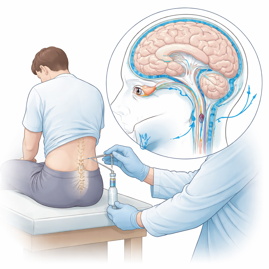
Lumbar puncture, often called a spinal tap, is a routine medical test used to diagnose infections, autoimmune diseases, and other brain or nerve problems. Yet a notable minority of patients develop a striking, posture‑dependent headache afterward: it eases when lying flat and worsens on sitting or standing. This study asks a practical question that matters to both patients and clinicians: can a quick ultrasound scan of the eye, done before and after the procedure, reveal who is at risk for this post‑procedure headache and help track their recovery?
Looking at Brain Pressure Through the Eye
The key idea behind the research is that the clear fluid surrounding the brain and spinal cord also extends along the optic nerve, the cable that carries visual signals from the eye to the brain. That nerve runs inside a sheath that can subtly widen or narrow as pressure in the head rises or falls. With gentle ultrasound over the closed eyelid, doctors can measure the diameter of this sheath in a few seconds. Earlier work showed that a wider sheath often signals high pressure in the skull; the team here explored the flip side: whether a shrinking sheath could signal low pressure and the special kind of headache that can follow a lumbar puncture.
How the Study Was Carried Out
Researchers at a university hospital in Germany followed 76 adult patients who needed a lumbar puncture for diagnosis only, not for anesthesia or surgery. All patients had normal scans of the head beforehand and no signs of dangerously raised pressure. Using a standardized ultrasound method, a trained examiner measured each patient’s optic nerve sheath diameter just before the puncture (T0), immediately afterward (T1), and again 24 hours later (T2). If a patient developed the characteristic postdural puncture headache—worse on standing, better when lying flat—extra measurements were taken at 48 and 72 hours. The team also collected age, sex, body mass index, needle size, and how much spinal fluid was removed.
What Happened to the Eye Measurements
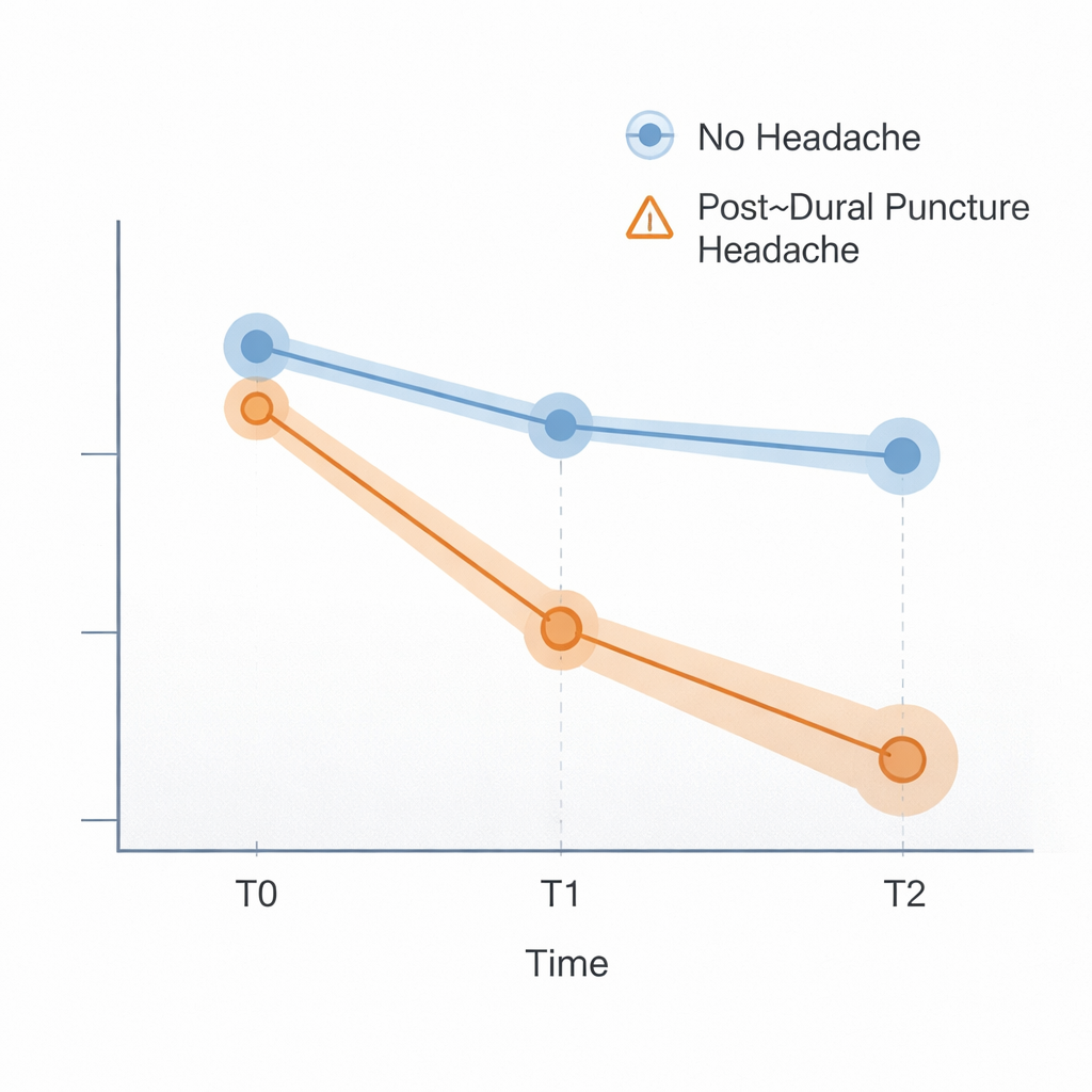
Every single patient showed a drop in optic nerve sheath diameter right after the lumbar puncture, confirming that removing spinal fluid causes a rapid, measurable fall in pressure transmitted up to the eye. Most patients’ measurements had bounced back close to their starting values by 24 hours. But in the seven patients—about 9 percent—who went on to develop the classic post‑procedure headache, the sheath remained clearly narrower at 24 hours and tended to shrink further by 48 hours. Statistical analysis showed that at the 24‑hour mark, the average sheath size differed sharply between those with and without headache, even though the two groups were similar in body build, sex, needle size, and amount of fluid taken. The only clear background difference was age: younger patients were more likely to get the headache.
A Cutoff Value and Its Promise
Using a type of analysis called a ROC curve, the authors sought a practical threshold: a sheath diameter small enough at 24 hours to flag a high risk of postdural puncture headache. They found that a value below 4.9 millimeters at that time point distinguished most affected patients from those without headache, with about 86 percent sensitivity (catching most true cases) and 93 percent specificity (few false alarms). While these numbers come from a modest sample, they suggest that a simple bedside eye scan could become part of a monitoring toolkit, especially for younger and otherwise higher‑risk patients after a spinal tap.
What This Means for Patients
For people undergoing a lumbar puncture, this work shows that a painless ultrasound of the eye can track how the pressure around the brain is changing in real time. A pronounced and lingering narrowing of the optic nerve sheath a day after the procedure appears to go hand in hand with the distinctive headache caused by low spinal fluid pressure. Although the study is relatively small and the cutoff value needs confirmation in larger, multi‑center trials, the approach offers a noninvasive way to spot trouble early and to follow recovery, potentially guiding decisions about rest, further imaging, or treatments such as an epidural blood patch.
Citation: Merzou, F., Kunzmann, AL., Janitschke, D. et al. Ultrasound measurement of optic nerve sheath diameter pre and post lumbar puncture for prediction of postdural puncture headache. Sci Rep 16, 7468 (2026). https://doi.org/10.1038/s41598-026-40311-1
Keywords: lumbar puncture, postdural puncture headache, optic nerve ultrasound, intracranial pressure, spinal tap complications