Clear Sky Science · en
Comparative evaluation of MRI-based bone-targeted sequences and computed tomography for preoperative assessment of midfacial trauma
Why safer face scans matter
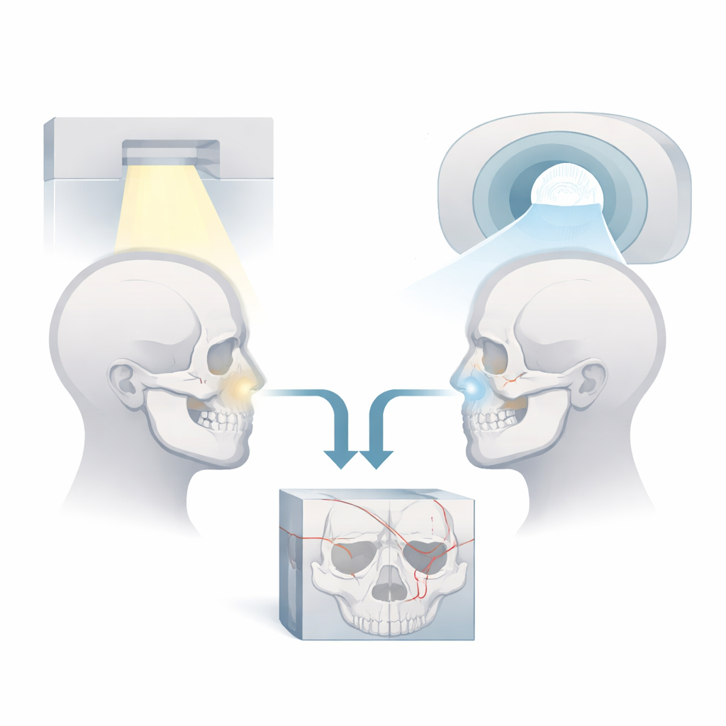
When someone is hit in the face in a car crash, a fight, or a sports accident, doctors must quickly see which delicate bones around the eyes, nose, and cheeks are broken. Today, this is usually done with a CT scan, which uses X‑rays and therefore exposes patients to radiation. This study asks a simple but important question: can modern MRI scans, which use magnets instead of X‑rays, give surgeons nearly the same detail for planning facial fracture surgery, while avoiding radiation—especially for younger and more vulnerable patients?
Looking inside the broken midface
The midface is a crowded crossroads of tiny bones, air spaces, nerves, and soft tissues around the nose, eye sockets, and upper jaw. In serious accidents, several of these structures can break at once, making it hard to judge the true extent of damage just by looking at the patient. CT scans are fast, widely available, and excellent at showing fine bony details, which is why they have long been the standard tool in emergency rooms. However, repeated CT scans add up to a higher lifetime radiation dose, which is a concern for children, young adults, and anyone who may need multiple scans over time.
A head‑to‑head test of CT and advanced MRI
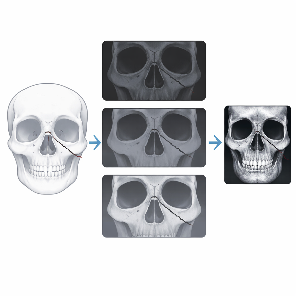
To compare these methods, the researchers followed 20 adults with fresh midface injuries—42 fractures in total. Every patient first received a CT scan, then a high‑resolution MRI exam on a powerful 3‑Tesla machine using a special coil designed to fit the jaw and face closely. The MRI session included five different "flavors" of 3D imaging sequences, each tuned in a different way to make bone stand out: UTE, DESS, Dark Bone, StarVIBE, and STIR. Three experienced readers—a radiologist and two maxillofacial surgeons—reviewed every CT and MRI dataset independently. They judged whether each fracture was correctly found and precisely located, and also rated image sharpness, visibility of fracture lines, clarity of bone edges, and contrast between bone and soft tissue.
How well MRI kept up with CT
CT lived up to its reputation, spotting 98% of all fractures with perfect agreement among readers and the shortest reading times, typically under a minute. MRI took a bit longer to interpret, but performance varied strongly between sequences. Two newer, gradient‑echo‑based techniques—called UTE and StarVIBE—came closest to CT. They detected about 88–89% of fractures, produced crisp images with excellent contrast, and showed high agreement between observers. These sequences were especially good at visualizing injuries of the eye socket, cheekbone complex, nasal bones, and walls of the maxillary sinus, where very thin bone must still be distinguished from nearby air and soft tissue. The other MRI sequences, particularly DESS and STIR, lagged behind, sometimes missing subtle or very fine fracture lines.
Where MRI shines and where CT still leads
Beyond simply seeing fractures, MRI has a natural advantage in showing the surrounding soft tissues, such as swollen muscles, trapped nerves, bleeding, and brain or eye complications. In several example cases, the best MRI sequences not only traced the broken bone but also revealed blood collections, sinus blockage, and possible injury to nearby coverings of the brain. On the other hand, MRI exams take longer to acquire, are more sensitive to patient motion, and still do not quite match CT in capturing every tiny crack in the most complex areas of the midface. For severely injured, unstable patients who need very rapid decisions, CT therefore remains the first‑line tool.
What this means for patients
Overall, the study shows that carefully chosen modern MRI techniques—especially UTE and StarVIBE—can come surprisingly close to CT in mapping midface fractures, while completely avoiding radiation and adding valuable soft‑tissue detail. The authors conclude that CT should still be used for urgent, high‑risk facial trauma and for the most intricate fracture patterns. But in more stable situations, for planned surgery, repeat follow‑up, or in younger and radiation‑sensitive patients, a tailored MRI‑based approach can serve as a realistic, safer alternative. In practical terms, this work brings doctors a step closer to planning facial fracture surgery with magnets instead of X‑rays, reducing long‑term risks without sacrificing crucial diagnostic information.
Citation: Al-Haj Husain, A., Kessler, P., Lie, S.A.N. et al. Comparative evaluation of MRI-based bone-targeted sequences and computed tomography for preoperative assessment of midfacial trauma. Sci Rep 16, 9700 (2026). https://doi.org/10.1038/s41598-026-40252-9
Keywords: midfacial fractures, CT-like MRI, radiation-free imaging, maxillofacial trauma, preoperative planning