Clear Sky Science · en
Ultra-high resolution multimodal MRI densely labelled holistic structural brain atlas
A New Map of the Living Human Brain
When doctors and scientists look at brain scans, they rely on reference maps—"atlases"—to understand what they are seeing. This paper presents a new kind of brain atlas, built from exceptionally sharp MRI scans and combining many pieces of expert knowledge into one detailed, easy-to-use map. It offers a clearer picture of the living human brain than most existing atlases and could help detect brain diseases earlier and study them more precisely. 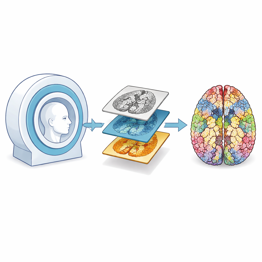
Why Brain Maps Matter
Brain atlases act like geographic maps for the mind. They divide the brain into recognizable regions, give each region a standard location, and make it possible to compare scans from different people or different hospitals. Today, such maps guide brain surgery, help researchers study how diseases like Alzheimer’s or Parkinson’s change brain structure, and support large-scale studies that combine data from many research groups. Most current atlases, however, are limited in detail and often focus on a single type of MRI image, which means subtle changes in small brain areas can be missed.
Building a Sharper Brain Picture
The authors created a new atlas, called holiAtlas, from MRI scans of 75 healthy young adults. Instead of the usual one-millimeter cubes used in standard brain maps, they worked at eight times finer resolution in each direction, allowing them to see much smaller features. They used three different types of MRI images, each emphasizing different tissues, and even generated a special kind of image—particularly good for deep brain structures—by training an artificial intelligence model on a separate dataset. All scans were carefully cleaned, aligned into the same space, and repeatedly averaged and refined so that the final images are both sharp and representative of a typical young adult brain.
Combining Many Expert Tools into One Atlas
To turn the images into a labeled map, the team did not start from scratch. Instead, they drew on seven state-of-the-art software tools, each specialized in particular brain regions, such as the cerebellum, hippocampus, hypothalamus, brainstem, or the folds on the brain’s surface. These tools automatically divided the brain into hundreds of subregions. The authors then corrected systematic mistakes—such as mislabeling blood vessels as brain tissue—using both advanced algorithms and careful human review. They resolved conflicts where different tools disagreed, applied anatomical rules to keep labels consistent, and filled in gaps so that every part inside the skull was meaningfully assigned. 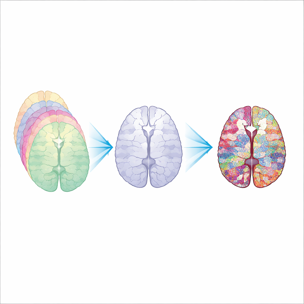
From Whole Brain to Tiny Parts
The finished atlas recognizes 350 tiny subregions, which can be grouped into 54 larger structures, then into nine broad tissue types, and finally into the entire intracranial volume. This "zoomable" structure lets users choose how much detail they need—from whole-brain studies to focused analysis of specific nuclei deep inside the brain. Crucially, all of these labels are based on what can actually be seen in standard, noninvasive MRI at 3-tesla field strength, making the atlas directly applicable in many hospitals and research centers without requiring exotic scanners or post-mortem tissue.
What This Could Mean for Brain Health
Because holiAtlas is sharper and more finely divided than most existing MRI-based maps, it may reveal small changes in particular subregions that signal disease much earlier. For example, only certain parts of the hippocampus and amygdala shrink early in Alzheimer’s disease, and specific deep nuclei are affected in Parkinson’s disease. Having a detailed, standardized map of these areas in a living, healthy brain provides a powerful reference for spotting such changes. The atlas is freely available, so it can be used to develop better analysis software, guide machine-learning tools, and support education—bringing us a step closer to understanding, and eventually treating, the brain’s many disorders.
Citation: Manjón, J.V., Morell-Ortega, S., Ruiz-Perez, M. et al. Ultra-high resolution multimodal MRI densely labelled holistic structural brain atlas. Sci Rep 16, 9457 (2026). https://doi.org/10.1038/s41598-026-40186-2
Keywords: brain atlas, MRI, neuroimaging, neurological diseases, brain mapping