Clear Sky Science · en
3D ultrasound assessment of the central sulcus in very preterm infants: feasibility and reproducibility of opening metrics study
Why tiny brain folds matter
Each year, thousands of babies are born far earlier than expected. Even when they survive the fragile first weeks of life, many face later challenges with movement, learning, and behavior. Doctors know these problems are linked to how the brain grows in the newborn intensive care unit, but they lack simple ways to watch that growth unfold day by day. This study explores whether a familiar bedside tool—ultrasound—can be upgraded to give a three-dimensional view of one key brain groove, the central sulcus, which helps control movement.
A closer look at an important brain groove
The central sulcus is a deep fold that separates the brain regions in charge of planning movement and sensing touch. It forms early in pregnancy and continues to change rapidly around the time very preterm infants are born. Earlier MRI studies have shown that the shape of this groove relates to later motor skills, but MRI scans are costly, require moving fragile babies out of the unit, and usually provide only a few time points. The authors asked whether three-dimensional ultrasound, performed through the soft spot on a baby’s head, could capture the size and shape of the central sulcus often enough and clearly enough to be useful for routine monitoring.
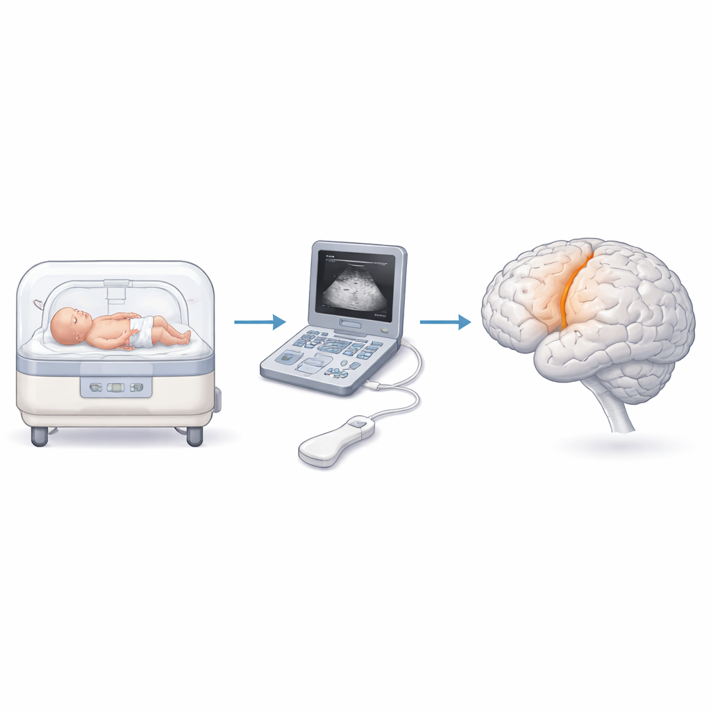
Turning bedside scans into 3D measurements
Thirteen very preterm infants, all born before 32 weeks of pregnancy, were followed in a single neonatal unit. At several points during their hospital stay, the team acquired 3D ultrasound volumes of each baby’s brain—109 scans in total, with three "sweeps" taken at each session. Using custom computer software, trained raters traced the central sulcus in these volumes and extracted twelve shape measurements, such as how long the sulcus was, how deep it reached, and how wide it opened along its course. The researchers then asked two questions: how consistent were these measurements when repeating scans on the same day, and did they change in a meaningful way as the babies grew older?
What the growing groove revealed
On single ultrasound sweeps, measurements varied quite a bit from one acquisition to the next, limiting their usefulness for one-off decisions about an individual child. However, when the three sweeps from the same day were averaged, the picture became much clearer. In particular, the average width, or "opening," of the sulcus could be measured with good reproducibility, approaching reliability levels considered acceptable for clinical tools. As expected for a structure that is still maturing, several features of the sulcus increased with the babies’ postmenstrual age: its length, its maximum depth, and the average opening all tended to grow between scans taken before 28 weeks and those closer to 36 weeks.

Right and left sides do not grow the same
By comparing the two brain hemispheres, the team also uncovered a clear asymmetry. Across the study, the right central sulcus consistently showed wider openings than the left. This rightward difference echoes findings from MRI work in larger groups of preterm and older children, and may be related to the early development of handedness and other forms of brain lateralization. The fact that bedside ultrasound could detect such subtle, side-to-side differences suggests that the technique is sensitive enough to track not only overall growth but also more nuanced patterns of brain shaping.
What this could mean for preterm babies
For families and clinicians, the main message is that 3D ultrasound can provide a window into how a vulnerable newborn’s brain folds and grows over time—without moving the baby, using radiation, or giving sedation. While this small pilot study cannot yet link specific ultrasound measurements to future motor or cognitive outcomes, it shows that averaging several bedside scans yields stable indicators of how the central sulcus is maturing. With larger studies and combined MRI data, such measures could help identify infants whose brain folds are not developing as expected, guide protective care strategies in the NICU, and perhaps one day refine early predictions of which children are at higher risk for movement and learning difficulties.
Citation: Barrios, C.R., Rosa, I.G., Fernández, S.P.L. et al. 3D ultrasound assessment of the central sulcus in very preterm infants: feasibility and reproducibility of opening metrics study. Sci Rep 16, 10199 (2026). https://doi.org/10.1038/s41598-026-40148-8
Keywords: preterm infants, 3D ultrasound, brain development, central sulcus, neonatal intensive care