Clear Sky Science · en
Machine-learning–guided transcriptomic integration identifies GFM1 as a lactylation-related candidate biomarker in aortic dissection
Why this hidden threat in the aorta matters
Aortic dissection is a medical emergency in which a tear inside the body’s main artery can cause life-threatening internal bleeding within hours. Doctors can often save patients with urgent surgery, but there are still no reliable blood tests to warn of danger early or drugs that slow the disease itself. This study explores whether subtle shifts in how artery cells handle energy and chemical signals might reveal new warning signs, focusing on a little-known gene called GFM1 that may help link cell metabolism to the weakening of the aortic wall.
Cracking the code of a dangerous tear
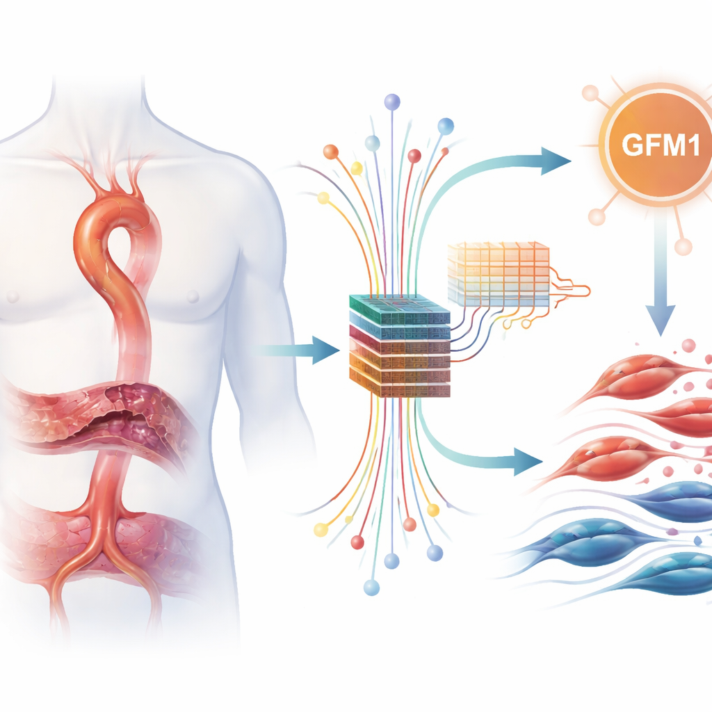
In aortic dissection, blood forces its way into the wall of the aorta, splitting its layers apart. Why some people’s aortas fail in this way is still not fully understood. The authors looked at patterns of gene activity in samples from people with aortic dissection and from individuals with healthy aortas. They paid special attention to genes tied to “lactylation,” a recently discovered way that cells use lactate—better known as the substance that builds up in muscles during hard exercise—to fine-tune proteins and gene regulation. Because lactylation has been linked to inflammation and tissue remodeling, the team suspected that lactate-related genes might also be involved in damaging the aortic wall.
Mining big data with smart algorithms
To test this idea, the researchers pooled several publicly available datasets that record which genes are turned up or down in aortic tissue. They carefully corrected for technical differences between studies and then searched for genes whose activity consistently differed between diseased and healthy samples. Out of thousands of genes, they found 217 with clear changes, many of which pointed to immune reactions and remodeling of the tissue scaffold that supports the aorta. Next, they focused on a curated list of genes related to lactate handling and lactylation and identified 11 that were both altered in aortic dissection and part of these lactate-linked programs.
Letting machines vote on a key suspect
Finding 11 interesting genes was still too many to study deeply in the lab, so the team turned to machine-learning methods as an objective “voting system.” They fed the data for these genes into three different models—LASSO, Random Forest, and XGBoost—that are commonly used to pick out patterns that best separate patients from controls. Each method highlighted its own favorites, but only one gene, GFM1, was strongly and consistently chosen by all three. This cross-checking approach made GFM1 stand out as a particularly robust candidate marker, even though the models were used for ranking rather than for building a ready-to-use diagnostic test.
Zooming in on artery muscle cells
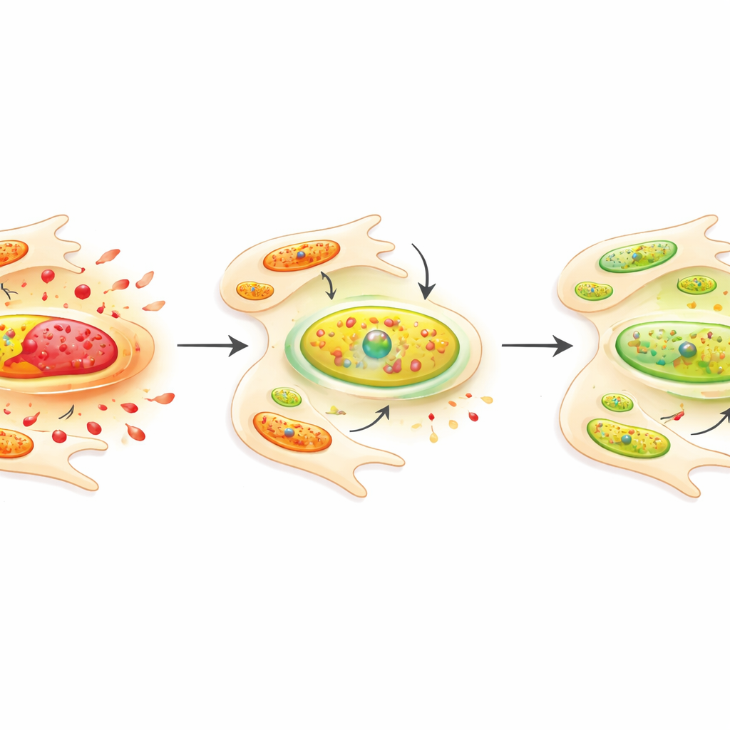
GFM1 helps control how mitochondria, the energy factories inside cells, build their own proteins. Because energy balance is crucial for how artery wall cells behave, the authors examined GFM1 more closely. They confirmed that GFM1 levels were higher in tissue from patients with aortic dissection than in non-diseased aortas. Then they moved to a controlled cell culture system using mouse vascular smooth muscle cells—the muscle-like cells that give the aorta its strength. When these cells were stimulated with angiotensin II, a hormone linked to high blood pressure and vessel stress, they became more prone to multiply and move, mimicking harmful changes seen in diseased arteries. When the researchers used small interfering RNA to lower GFM1 in these cells, the angiotensin-driven growth and migration were noticeably reduced, suggesting that GFM1 helps promote these risky behaviors.
What this means and what it does not yet prove
Taken together, the findings suggest that GFM1 may act as a bridge between altered cell metabolism and the aggressive behavior of artery wall cells in aortic dissection. In simple terms, higher GFM1 activity appears to go hand in hand with a more unstable, damage-prone aorta, and dialing it down in lab-grown cells makes them less likely to overgrow and migrate. However, the authors are careful to stress that this work is still at an early, hypothesis-building stage. They did not directly measure lactylation in tissues or prove that GFM1 itself is chemically modified in this way, and the predictive power of the models has not been tested in independent patient groups. Future studies will need to confirm these results in larger cohorts and explore exactly how GFM1 and related metabolic changes weaken the aortic wall. If those efforts succeed, GFM1 or its pathways could eventually become targets for new blood tests or treatments aimed at preventing this often fatal tear before it occurs.
Citation: Chen, J., Jiang, N., Guo, Z. et al. Machine-learning–guided transcriptomic integration identifies GFM1 as a lactylation-related candidate biomarker in aortic dissection. Sci Rep 16, 9033 (2026). https://doi.org/10.1038/s41598-026-40139-9
Keywords: aortic dissection, vascular smooth muscle cells, lactate metabolism, mitochondrial function, biomarker discovery