Clear Sky Science · en
Development of a CT radiomics and clinical feature combined model for predicting early recurrence of surgical resected hepatocellular carcinoma
Why this matters for people with liver cancer
For many people with liver cancer, surgery offers the hope of cure—yet the cancer often comes back within just a couple of years. This study asks a crucial question: can we use information already hidden in routine medical scans and common lab tests to spot, before surgery, who is most likely to see their cancer return early? If so, doctors could tailor follow-up care and extra treatments to those who need them most.
Seeing more inside standard CT scans
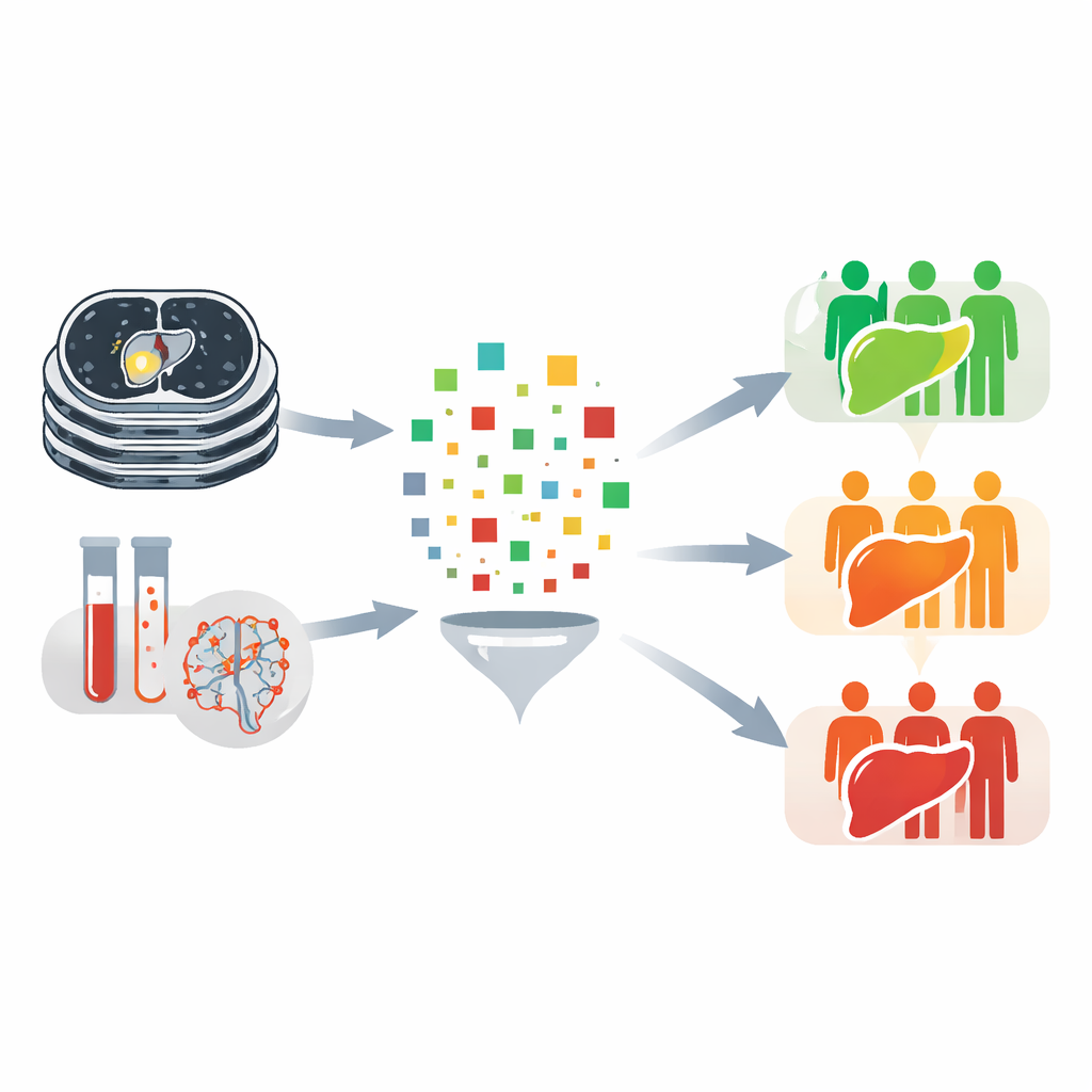
People with hepatocellular carcinoma, the most common type of liver cancer, usually undergo contrast-enhanced CT scans before surgery. Radiologists look at these images to judge tumor size, number, and location. But computers can go further, measuring subtle patterns of brightness, shape, and texture that our eyes cannot easily discern. In this study, the researchers analyzed pre-surgery CT scans from 436 patients who had their liver tumors removed. Using specialized software, they outlined each person’s main tumor in three dimensions and automatically extracted almost two thousand numerical features describing the tumor’s appearance in two contrast phases.
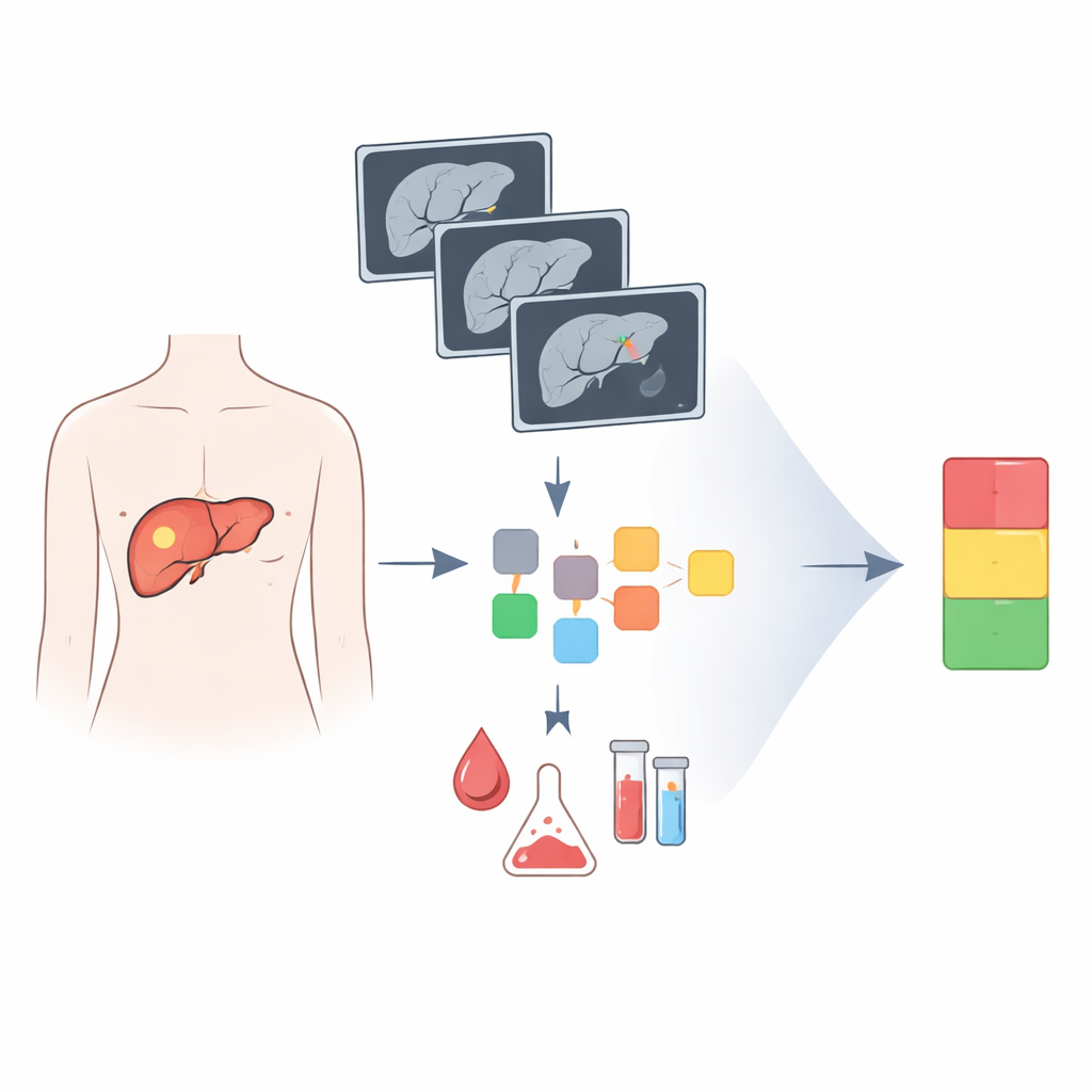
Building a risk score from images and blood tests
From this large pool of imaging features, the team applied a statistical method designed to keep only the most informative signals while discarding noise. They ended up with 20 CT-based features that were most strongly linked to whether a patient’s cancer returned within two years after surgery. These features were combined into a single "radiomics" score for each person. The researchers then examined many clinical factors—such as tumor size, blood test results, and microscopic signs of tiny vessel invasion in the removed tissue—to see which best predicted early return of cancer.
A simple tool to sort patients into risk groups
Four factors emerged as the most powerful predictors when considered together: the CT-based radiomics score, the presence of cancer cells in small blood vessels near the tumor, the level of a liver enzyme relative to immune cells in the blood, and the level of a common tumor marker measured in blood. The authors combined these into a visual scoring tool that outputs each patient’s likelihood of remaining free of recurrence at two years. When they tested this tool in one group of patients and then checked it in a separate group, it reliably separated people into low-, intermediate-, and high-risk categories, with clearly different chances of remaining cancer-free. Importantly, the model also worked well in patients whose usual tumor marker is not elevated, a group for whom prediction has been especially challenging.
Linking scan patterns to tumor biology
To explore why the CT-based score might be so informative, the researchers looked at tumor samples from a smaller subset of patients and measured the activity of eleven genes previously tied to aggressive liver cancer. They found that higher radiomics scores were moderately associated with increased activity of two genes that have been implicated in tumor growth and spread. Although early and exploratory, this suggests that the patterns computers detect on CT images may reflect underlying genetic programs that drive more dangerous behavior, offering a window into tumor biology without the need for repeated biopsies.
What this could mean for care after surgery
Overall, this work shows that routine CT scans and common lab tests, when analyzed with modern computational methods, can help forecast which liver cancer patients are most likely to face an early return of their disease after surgery. The resulting risk groups could guide how closely patients are monitored and who might benefit from additional treatments soon after their operation. While the model needs to be confirmed in larger and more diverse groups and its biological underpinnings better understood, it represents a step toward more personalized follow-up plans that match the intensity of care to each patient’s actual risk.
Citation: Liao, M., Liao, N., Huo, S. et al. Development of a CT radiomics and clinical feature combined model for predicting early recurrence of surgical resected hepatocellular carcinoma. Sci Rep 16, 10453 (2026). https://doi.org/10.1038/s41598-026-40130-4
Keywords: liver cancer, CT imaging, radiomics, cancer recurrence, risk prediction