Clear Sky Science · en
Ciprofol attenuates cerebral Ischemia‒reperfusion injury in rats by inhibiting ferroptosis through upregulating AMPK
Why Protecting the Brain After Stroke Matters
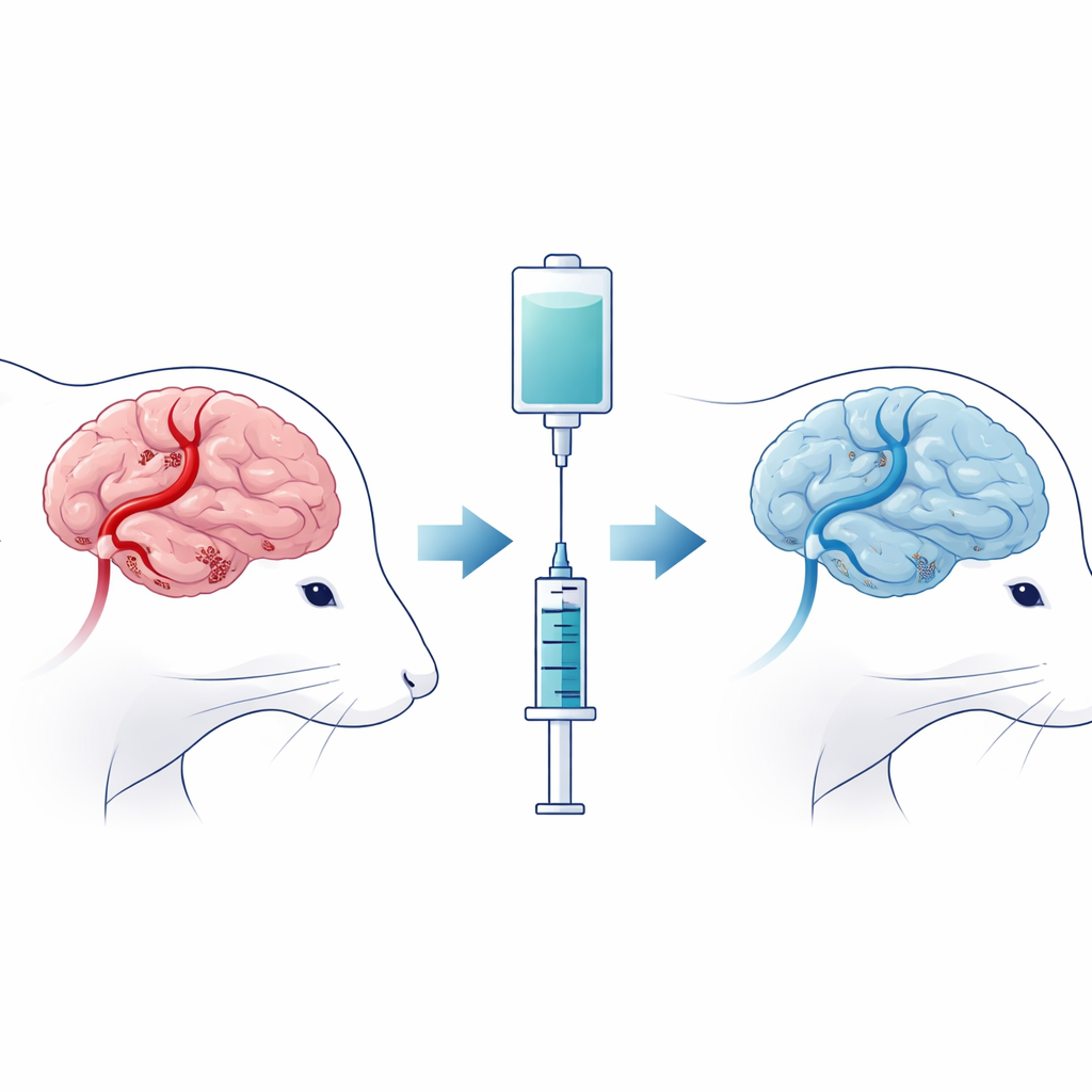
When a person has an ischemic stroke, doctors race to reopen the blocked blood vessel and restore blood flow to the brain. Paradoxically, this life‑saving step can itself cause extra damage, a phenomenon called reperfusion injury. The brain, suddenly flooded with oxygen and nutrients after a period of deprivation, unleashes a storm of chemical reactions that can kill vulnerable nerve cells. This study explores whether ciprofol, a new anesthetic drug already used for sedation, can also act as a protector for the brain during this critical window by calming some of the most harmful processes triggered after blood returns.
From Blocked Blood Flow to Brain Damage
To mimic what happens in a human stroke, the researchers temporarily blocked a major brain artery in rats and then restored blood flow, creating what is known as cerebral ischemia–reperfusion injury. Some rats simply underwent the procedure, while others received ciprofol shortly after blood flow was restarted. The team then assessed how well the animals could move and respond to touch, and examined their brains for regions of dead tissue and structural damage in nerve cells. Rats that received ciprofol had smaller areas of brain injury, healthier‑looking nerve cells, and better movement and sensory scores than untreated animals, suggesting that the drug blunted the worst consequences of the insult.
A Hidden Form of Cell Death Driven by Iron
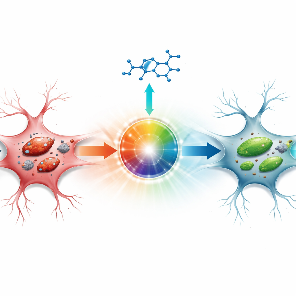
Beyond visible tissue damage, the authors focused on a recently recognized type of cell death called ferroptosis, which is fueled by iron and runaway chemical reactions that attack the fatty components of cell membranes. In untreated stroke‑model rats, brain tissue from the vulnerable zone around the core of the injury contained more iron, higher levels of a lipid damage by‑product, and nerve cell mitochondria that looked swollen and structurally broken under the electron microscope. Molecular tests showed that protective proteins that normally detoxify harmful lipid by‑products were reduced, while proteins that promote this destructive process were increased. Ciprofol largely reversed these changes: iron and lipid damage fell, the balance of key proteins shifted toward protection, and mitochondria maintained more normal shapes and internal structure.
A Cellular Energy Sensor as the Control Switch
The study then probed how ciprofol produces these protective effects. Attention centered on AMPK, a protein that acts as a cellular energy sensor and stress responder. In the injured brains of untreated rats, AMPK activity was depressed. Ciprofol boosted the activated form of AMPK, in parallel with the reductions in ferroptosis‑related damage. To test whether this energy sensor truly sits upstream of the protective chain of events, the researchers used another drug, Compound C, that blocks AMPK. When animals received both ciprofol and this AMPK blocker, the benefits of ciprofol were partially lost: ferroptosis‑related markers crept back toward harmful levels, iron accumulation returned, and the signal of AMPK activity dropped even lower than in untreated stroke animals. This pattern supports the idea that turning on AMPK is a key step in how ciprofol shields brain cells.
Dialing Down the Brain’s Inflammatory Storm
Stroke‑related injury is not only a matter of cell death inside neurons; the brain’s immune response also plays a major role. The team measured several inflammatory messenger molecules that typically surge after ischemia–reperfusion. In untreated animals, these signals were strongly elevated, reflecting an intense inflammatory reaction. Ciprofol reduced all three major inflammatory markers measured, suggesting that it not only suppresses iron‑driven membrane damage but also tempers the inflammatory storm that follows. When AMPK was blocked with Compound C, these calming effects on inflammation were weakened, again tying the benefits of ciprofol to this central energy‑sensing pathway.
What This Could Mean for Future Care
Taken together, the results point to ciprofol as more than just a sedative: in this rat model, it appears to limit stroke‑related brain damage by activating AMPK, which in turn curbs iron‑driven cell death and dampens inflammation. Because ciprofol is already being used in clinics for anesthesia and procedural sedation, its added potential as a brain‑protective agent is especially intriguing. Still, the work has important limitations, including reliance on a single animal model and the possibility that the AMPK‑blocking drug has side effects of its own. More studies, including in other species and ultimately in human patients, will be needed to confirm whether ciprofol can safely help protect the brain during and after stroke treatment.
Citation: Zeng, H., Yu, X., Zheng, Z. et al. Ciprofol attenuates cerebral Ischemia‒reperfusion injury in rats by inhibiting ferroptosis through upregulating AMPK. Sci Rep 16, 9282 (2026). https://doi.org/10.1038/s41598-026-40104-6
Keywords: ischemic stroke, reperfusion injury, ciprofol, ferroptosis, neuroprotection