Clear Sky Science · en
Feasibility and histological analysis of multi-hole versus fully covered self-expandable metallic stents in a porcine model of hilar biliary obstruction
Why blocked bile ducts matter
When the tiny tubes that drain bile from the liver become squeezed shut, waste products build up in the body. People can develop yellowing of the skin, infections, pain, and even liver damage. Treating these narrowings, especially where many small bile ducts branch together high in the liver, is tricky. This study tested a new type of metal tube, or stent, in pigs to see whether it could keep bile flowing more naturally and irritate the liver less than the standard design.
A bottleneck at the fork in the road
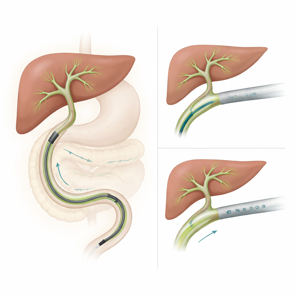
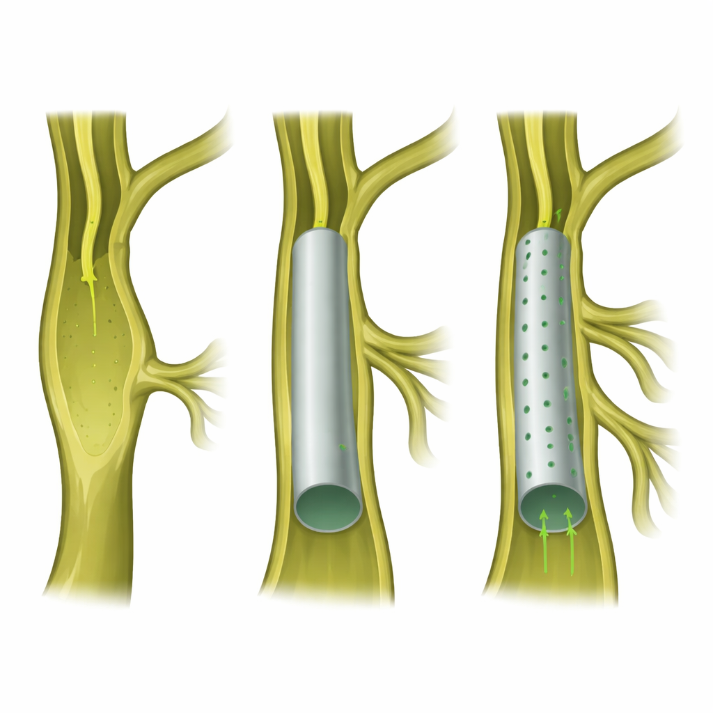
Near the center of the liver, major bile ducts split into many smaller branches, like a tree trunk dividing into limbs. Tumors or scarring in this region can pinch off the main channel, backing up bile into the liver. Doctors now often relieve this by sliding a stent into the duct through an endoscope passed from the mouth into the intestines. Traditional fully covered metal stents open the main passage well, but because they form a smooth tube, they can block off side branches, much like putting a sleeve over the trunk that also covers several limbs. That blockage can spark infection and further liver injury.
A metal tube with tiny escape routes
The new device, called a multi-hole self-expandable metallic stent, keeps the same basic metal mesh and protective coating as a standard covered stent but adds a row of small round openings along its length. These openings are designed to let bile trickle in and out of nearby side branches while still maintaining a wide main channel and resisting movement. Because it is fully covered, the stent is intended to be removable, allowing doctors to adjust or replace it if the patient’s condition changes over time.
Testing the idea in pigs
To study the stent safely and in detail, the researchers created controlled narrowings of the main bile duct near the liver in eight miniature pigs using a heat-based catheter. After the scars had time to form, each animal received either the new multi-hole stent or a standard fully covered stent placed through an endoscope. The pigs were then followed for three months with blood tests, X-ray dye studies of the bile ducts, and finally detailed microscope examination of the liver tissue around undrained side branches. The team focused on whether the stents stayed in place, could be removed, and how much inflammation, scarring, and damage appeared in side ducts that lay next to but not directly inside the stented segment.
What the stents did to the liver
All surviving pigs in both groups had their stents successfully placed and later removed using an endoscope. Two animals, one in each group, died from accidental perforations during the imaging step before stent insertion, not from the stents themselves. Among the remaining six, bile tests showed a pattern: animals with the multi-hole stent tended to have lower levels of bilirubin, a pigment that rises when bile cannot drain, while two of the three pigs with the standard stent briefly developed higher levels. Under the microscope, tissue near undrained branches in the multi-hole group generally showed milder inflammation and fibrosis than in the conventional group. Because there were only three animals per group, these differences did not reach the strict statistical thresholds scientists usually require, but the trends pointed in the same direction.
What this could mean for patients
For people with complex blockages high in the bile ducts, doctors face a trade-off: stents that are easy to remove and resist tissue ingrowth may also block important side branches and raise the risk of infection. This animal study suggests that a redesigned covered metal stent with small side openings may ease that trade-off by helping preserve drainage from more of the liver while remaining removable. The work does not yet prove that the new stent is better for patients with cancer or other diseases, and the pig model cannot fully mimic human tumors. Still, the safety of placement and removal, combined with signals of less tissue damage, support moving to larger human trials to find out whether this design can improve outcomes for people living with serious bile duct obstruction.
Citation: Kim, E.J., Kang, H., Park, J.K. et al. Feasibility and histological analysis of multi-hole versus fully covered self-expandable metallic stents in a porcine model of hilar biliary obstruction. Sci Rep 16, 9737 (2026). https://doi.org/10.1038/s41598-026-40067-8
Keywords: biliary stent, hilar biliary obstruction, liver drainage, endoscopic therapy, porcine model