Clear Sky Science · en
Brain tumor classification using optimized ResNet50 with dynamic precision optimization for enhanced speed and diagnostic accuracy
Smarter Scans, Faster Answers
Brain tumors are among the most frightening diagnoses a person can face, and every hour saved in finding and classifying them can matter. This study presents a new artificial intelligence (AI) system that reads brain MRI scans with near-perfect accuracy while using less computing power than many existing methods. That mix of speed, precision, and efficiency could help bring advanced diagnostic support not only to major hospitals, but also to clinics with more modest hardware. 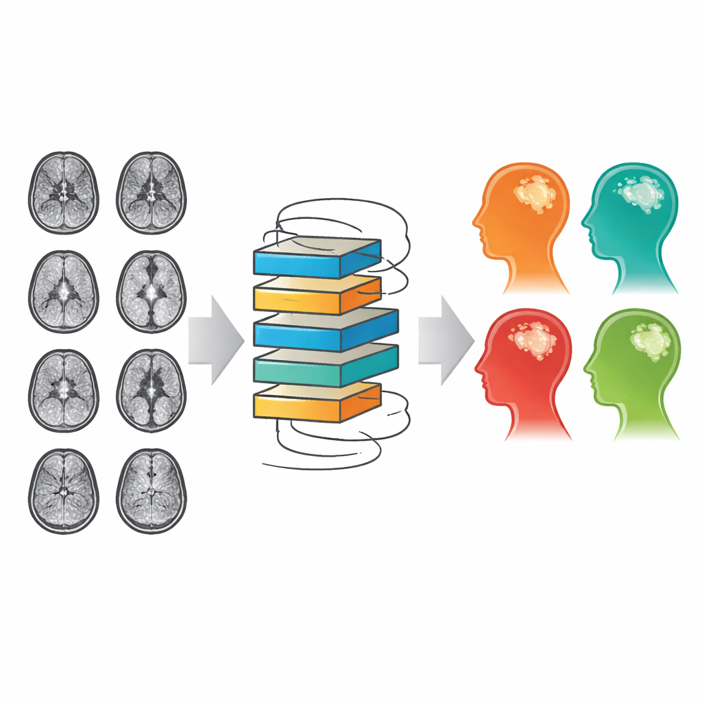
Why Brain Tumor Detection Is So Hard
Brain tumors come in many shapes, sizes, and locations, and even experts can struggle to distinguish subtle differences on MRI scans. The skull is a closed, rigid space, so any abnormal growth can disrupt vital brain functions, making early and accurate diagnosis essential. MRI is the imaging tool of choice because it provides detailed pictures of soft tissue without harmful radiation. But as datasets grow and tumor types become more finely classified, radiologists face an overwhelming number of images to inspect. This has fueled interest in computer systems that can automatically flag and classify tumors, helping doctors work faster and catch details that might otherwise be missed.
Building on a Proven AI Workhorse
The researchers started with ResNet50, a widely used deep learning model that has excelled at recognizing patterns in everyday photographs. ResNet50 is popular because its special “shortcut” connections allow it to be very deep without becoming unstable during training. However, the standard version is designed for three-color images and large datasets, and it consumes a lot of memory—problems for grayscale MRI scans and typical hospital hardware. The team adapted the first layer of ResNet50 to accept single-channel MRI images directly and replaced the bulky, general-purpose output layer with a lighter, task-specific classifier tuned to four categories: glioma, meningioma, pituitary tumor, and no tumor.
Doing More With Less Computation
To make the system both fast and accurate, the authors introduced a dynamic precision method that decides, on the fly, how carefully each part of the network needs to do its math. Most of the heavy image-processing layers run with lower-precision numbers that are faster and use less memory, while sensitive steps such as normalization and final decisions use full precision for stability. They also use transfer learning, which means the model reuses knowledge learned from millions of general images and then fine-tunes itself on a smaller brain MRI dataset. Data augmentation—simple flips, rotations, and brightness changes—further teaches the network to recognize tumors even when scans vary slightly. Together, these steps trimmed the number of parameters by about 3.7%, cut training time by more than 12%, and reduced graphics memory usage by over 40% without sacrificing performance. 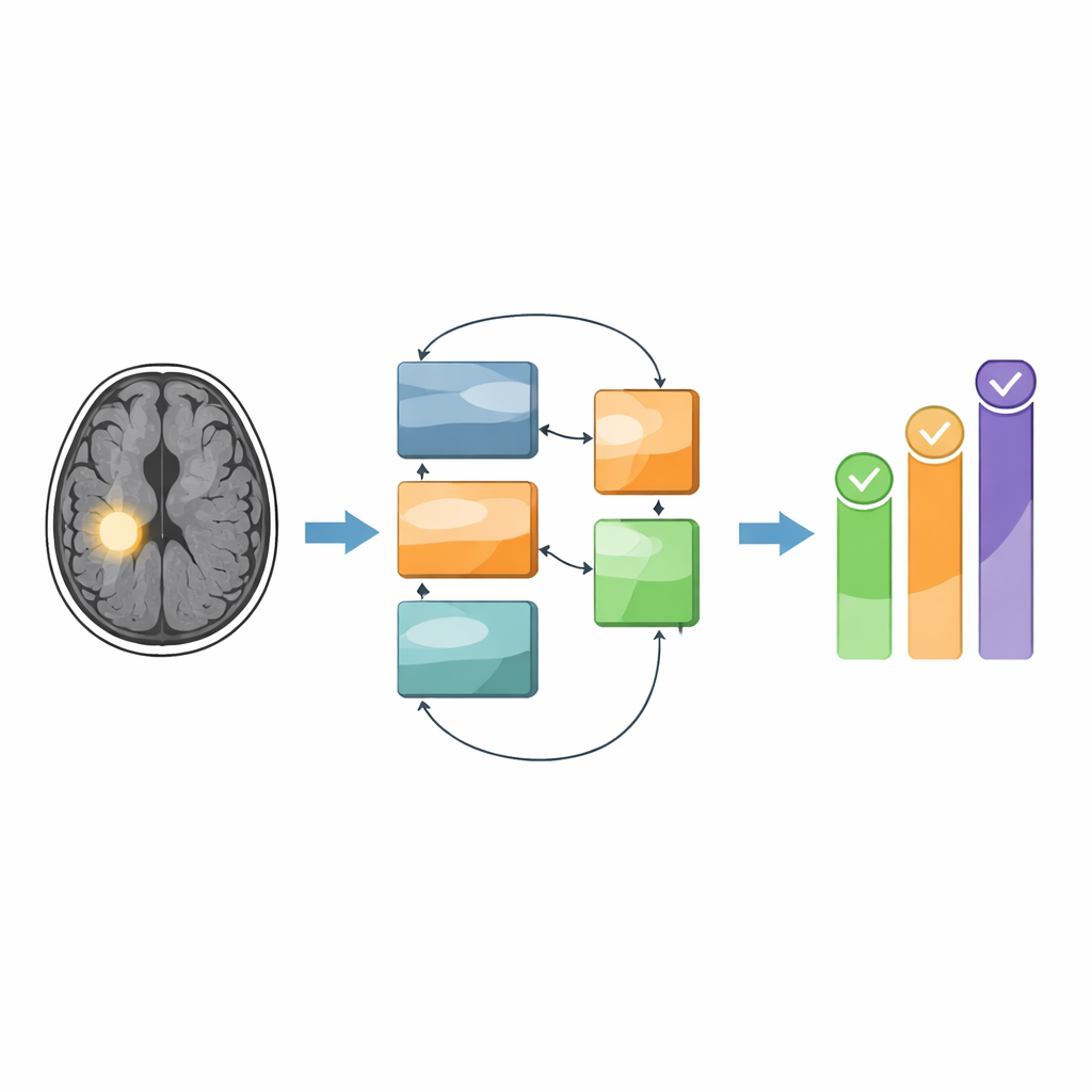
Making AI Decisions Easier to Trust
High accuracy alone is not enough in medicine; doctors also need to understand why an AI system reached a particular conclusion. To address this, the researchers built a second, "hybrid" version of their system. In this setup, ResNet50 acts as a feature extractor, turning each MRI into a detailed numerical fingerprint. Instead of sending that directly to a typical deep learning output layer, they feed it into a Random Forest, a classic machine learning method made of many decision trees. This approach makes it possible to rank which features influence each decision and to generate visual maps that show which regions of the brain the network focused on. In tests, this hybrid system achieved 99.31% accuracy—slightly below the pure deep learning model but with the advantage of clearer, more traceable reasoning.
Performance That Rivals More Complex Models
The team evaluated their methods on a public collection of 7,023 MRI images drawn from three established datasets and split into four classes. The optimized ResNet50 reached an overall accuracy of 99.69%, correctly classifying almost every tumor and non-tumor case. It achieved 100% precision for glioma, pituitary, and healthy scans, and nearly perfect scores for meningioma. Detailed tests showed high sensitivity and specificity for each class, meaning the model was both good at catching true tumors and at avoiding false alarms. When compared against many recent approaches—including deeper networks and sophisticated hybrid schemes—the optimized ResNet50 either matched or outperformed them, all while using fewer parameters and running efficiently on standard graphics cards.
From Research to the Radiology Suite
The authors envision their system as a decision-support tool integrated into hospital imaging workflows rather than a replacement for radiologists. In practice, MRI scans would flow from existing hospital systems into the AI model, which would quickly propose a tumor category and highlight key regions of interest. Radiologists would then review these suggestions alongside the raw images, combining human judgment with machine speed. The study acknowledges that more work is needed, especially testing on larger and more diverse, multi-center datasets and incorporating other imaging methods. Still, the results suggest that carefully designed, resource-aware AI can provide fast, accurate, and interpretable help in diagnosing brain tumors, potentially improving care even in settings where computing power is limited.
Citation: Mehrdad, V., Talebzadeh, R. & Fazaeli, N. Brain tumor classification using optimized ResNet50 with dynamic precision optimization for enhanced speed and diagnostic accuracy. Sci Rep 16, 9263 (2026). https://doi.org/10.1038/s41598-026-39926-1
Keywords: brain tumor MRI, deep learning diagnosis, ResNet50 optimization, medical image AI, tumor classification