Clear Sky Science · en
Parameters associated with the size of the foveal avascular zone in healthy elderly eyes
Why the Center of Your Vision Matters
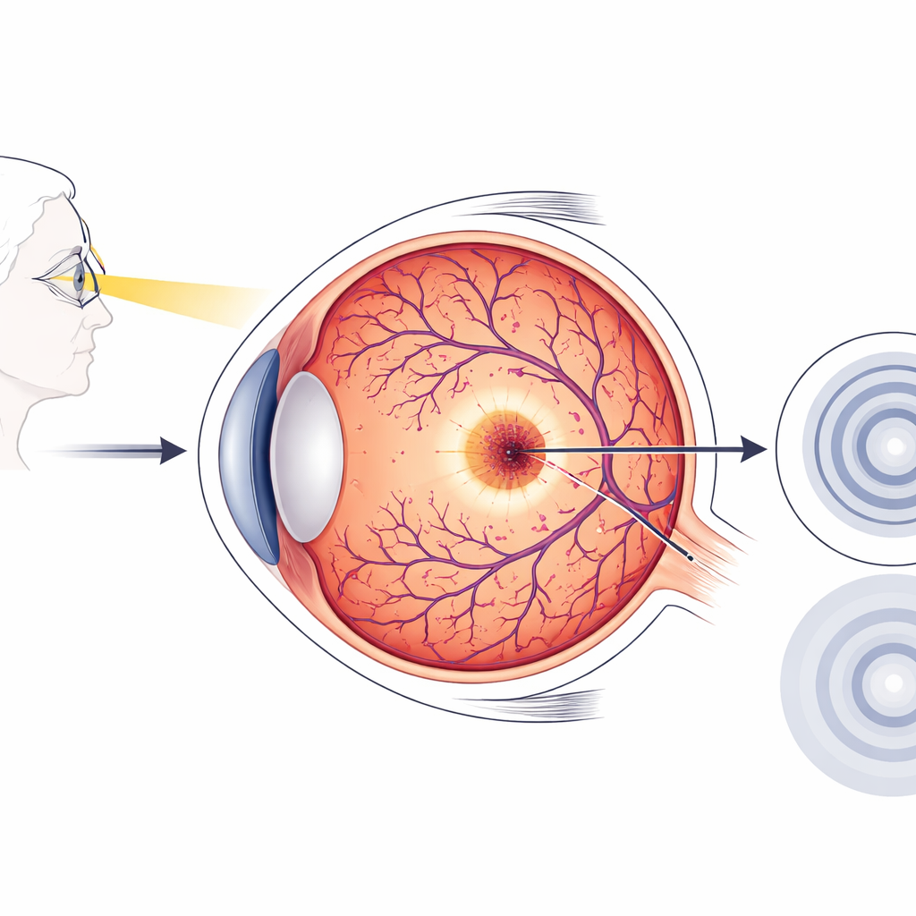
The tiny spot you use to read, recognize faces, or thread a needle sits at the very center of your retina and is called the fovea. To work at peak performance, this region must have a precise arrangement of light-sensing cells and blood vessels. This study looks closely at how that arrangement changes in healthy older adults, offering clues about normal aging in the eye and how to spot very early signs of disease.
A Closer Look at the Eye’s Sharpest Point
In the fovea, vision is sharpest because cone photoreceptors—cells that detect fine detail and color—are packed tightly together. Unusually, this central zone has no tiny blood vessels running through it; this empty ring is called the foveal avascular zone, or FAZ. Doctors can now map both the FAZ and the underlying light-sensing cells in fine detail using optical coherence tomography angiography, a noninvasive imaging method that produces detailed, map-like pictures of the retina’s blood supply and structure.
What the Researchers Wanted to Find
The team studied 101 eyes from people aged 50 and older who had excellent vision and no detectable retinal disease. They measured the area of the FAZ and how round it was, the thickness of the central retina, and the exact positions of two key landmarks: the center of the FAZ and the center of a small “bulge” in the layer of photoreceptors that marks where these cells are longest and most concentrated. By comparing the distance and direction between these two centers, the researchers asked whether subtle shifts in alignment were linked to how large the vessel-free zone becomes with age.
Subtle Shifts in Alignment With Age
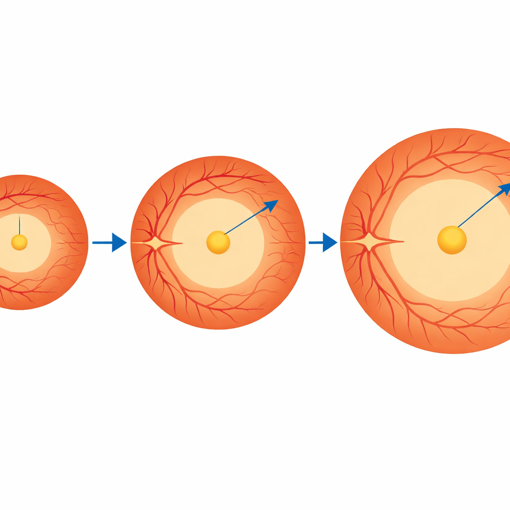
On average, the vessel-free zone in these healthy older eyes was about one-third of a square millimeter in area, but there was considerable natural variation from person to person. The central retina was thinner when the FAZ was larger, and thicker when the FAZ was smaller, reflecting how the foveal “pit” shape relates to the vessel-free area. Surprisingly, the overall distance between the centers of the FAZ and the photoreceptor bulge did not predict how large the FAZ was. What mattered instead was direction: in eyes where the FAZ lay more toward the temple side and slightly upward from the photoreceptor center, the vessel-free zone tended to be larger.
Clues From Development and Aging
Why would the vessel-free zone drift in a preferred direction? The authors suggest that both early development and lifelong blood flow patterns may play a role. During fetal growth, blood vessels approach the future fovea in an uneven way, especially on the temple side, which could set up a slight offset from the start. Later in life, differences in blood flow and oxygen demand between regions of the macula—such as the upper versus lower halves—may gradually reshape the fine network of capillaries. Over the years, these processes could nudge the FAZ outward in certain directions more than others, even in eyes that remain functionally normal.
What This Means for Aging Eyes
The study concludes that in healthy older adults, larger vessel-free zones are not simply scaled-up circles centered perfectly on the point of sharpest vision. Instead, they tend to expand more toward the upper and temple sides of the macula. Mapping not just the size but also the direction of this offset could help eye specialists distinguish normal aging patterns from the earliest changes of diseases such as diabetic retinopathy or macular degeneration. In everyday terms, understanding exactly how the “no-vessel” zone around our visual sweet spot shifts with age may give doctors a more sensitive early warning system for problems that threaten central vision.
Citation: Hasegawa, T., Nonaka, K., Onishi, T. et al. Parameters associated with the size of the foveal avascular zone in healthy elderly eyes. Sci Rep 16, 8064 (2026). https://doi.org/10.1038/s41598-026-39865-x
Keywords: foveal avascular zone, macular aging, retinal blood flow, optical coherence tomography angiography, central vision