Clear Sky Science · en
Three‑dimensional immune cartography uncovers subclinical remodeling in psoriasis
Why hidden changes in skin matter
People with psoriasis usually notice the condition only when red, scaly patches appear on their skin. But long before these plaques are visible, the immune system is already quietly reshaping the skin. This study uses a kind of three-dimensional "map" of skin samples to reveal how immune cells move and cluster beneath the surface, even in skin that still looks normal. Understanding these early, hidden changes could help doctors detect psoriasis activity sooner and design better targeted treatments.
Looking at skin in three dimensions
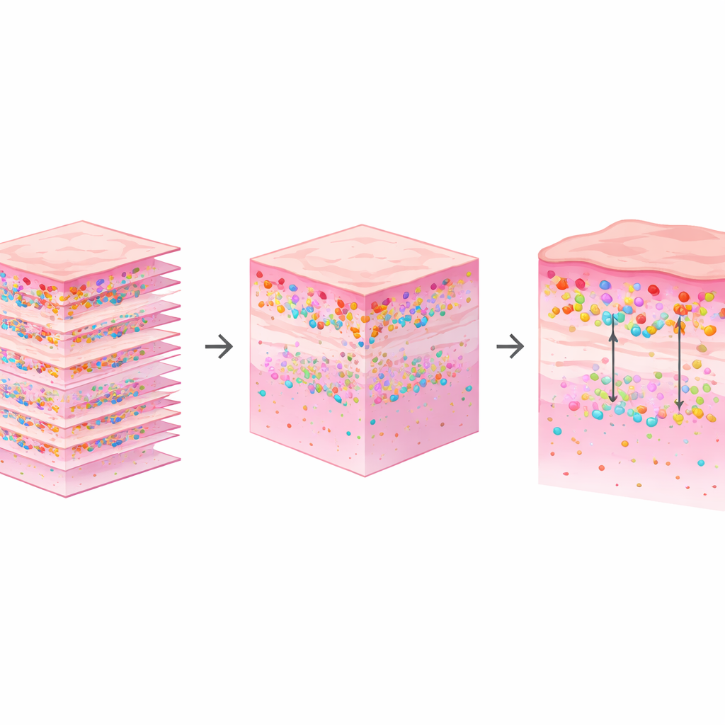
Most routine skin analysis relies on thin two-dimensional slices viewed under a microscope. While useful, this approach flattens what is in reality a three-dimensional landscape. The researchers set out to rebuild that third dimension using standard pathology methods already common in hospitals. They collected hundreds of very thin slices from preserved skin samples taken from people with psoriasis and from healthy volunteers. Each slice was stained so that key immune cells—T cells, macrophages, and mast cells—could be seen, then scanned into a computer. Using image-alignment algorithms and machine-learning-based tissue segmentation, the team digitally stacked these slices into full 3D blocks of skin, allowing them to see where different cells actually sit in depth.
How immune cells rearrange around psoriasis plaques
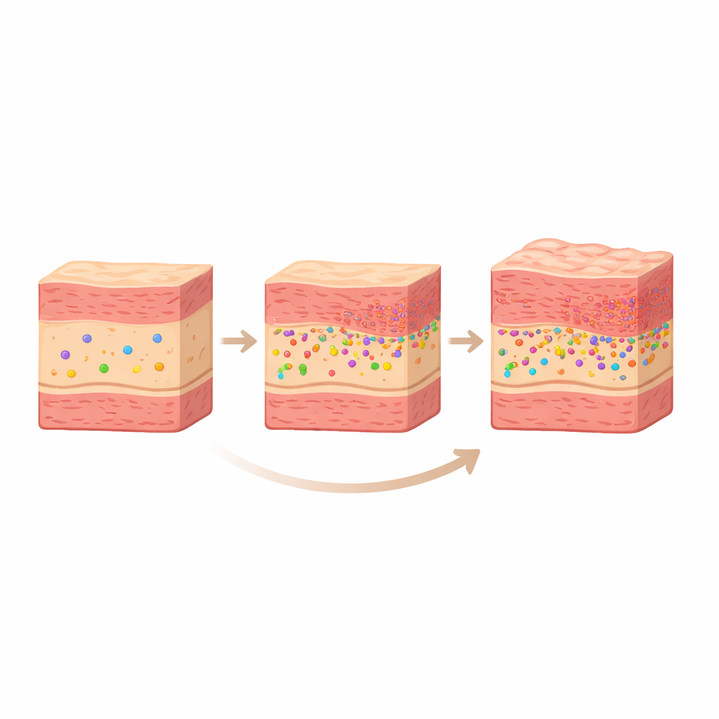
The team focused on three groups of skin: clearly diseased plaques, nearby normal-appearing skin within one centimeter of a plaque (called peri-lesional), and skin from people without psoriasis. For T cells, they found that in healthy skin these cells tend to sit deeper in the dermis, farther from the boundary where the inner skin meets the surface layer. In peri-lesional and especially in plaque skin, T cells shifted upwards, clustering much closer to this boundary, right under the outer skin cells that overgrow in psoriasis. Interestingly, the density of T cells dipped in peri-lesional skin compared with healthy skin, then rose again in full plaques, suggesting that T cells are not just increasing or decreasing in number; they are moving and redistributing as the disease evolves.
Early warning signs from macrophages and quiet mast cells
Macrophages, another major immune cell type marked here by CD68, also showed a meaningful shift. In plaque skin they became both more numerous and closer to the surface boundary than in healthy controls. Even in peri-lesional skin that looked normal to the naked eye, these cells were already positioned closer to that boundary than in healthy volunteers, hinting at a "primed" state of inflammation before plaques fully form. In contrast, mast cells behaved differently. Their overall numbers and average depth did not differ significantly among healthy, peri-lesional, and plaque samples. Subtle trends suggested that mast cells might move from deeper to more superficial zones as plaques mature, but the main message is that where mast cells sit may matter less than how activated they are—something this study’s staining approach cannot directly measure.
Why 3D views beat flat snapshots
A key technical insight of this work is that averaging many flat slices can actually hide important spatial patterns. When the researchers compared distance profiles calculated in true 3D with those obtained by averaging separate 2D slices, the 2D view smoothed over peaks and valleys in immune-cell distribution. No single slice accurately represented the full 3D pattern. Only by reconstructing the full volume could they clearly see how T cells and macrophages form layered clusters near the surface as psoriasis advances. This demonstrates that the skin’s immune landscape is not uniform in depth, and that volumetric analysis is better suited for capturing such structure.
What this means for people with psoriasis
In simple terms, the study shows that psoriasis is not just about the visible plaque; the surrounding skin is already subtly rewired by the immune system in three dimensions. T cells and macrophages quietly move closer to the surface long before dramatic scaling appears, while mast cells seem to play a more functional than positional role. By turning routine pathology slides into 3D immune maps, this accessible method adds depth—literally—to our understanding of psoriatic skin. In the future, such mapping could help link molecular readouts to physical cell locations, improve how we track disease activity, and inform therapies aimed at calming inflammation before it becomes visible at the surface.
Citation: Li, L., Vu, L., Drury, P. et al. Three‑dimensional immune cartography uncovers subclinical remodeling in psoriasis. Sci Rep 16, 10241 (2026). https://doi.org/10.1038/s41598-026-39838-0
Keywords: psoriasis, skin inflammation, immune cells, 3D imaging, digital pathology