Clear Sky Science · en
WISeRKNet: wide slice residual Kronecker network for lung cancer detection based on CT images
Why this matters to everyday health
Lung cancer remains one of the deadliest cancers worldwide largely because it is often discovered too late. This study explores how advanced computer techniques can help doctors find signs of lung cancer earlier and more accurately in common medical scans, potentially leading to faster treatment and better chances of survival.
Seeing inside the chest with clearer scans
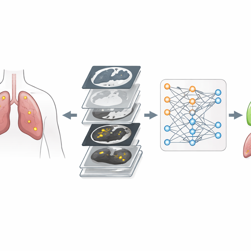
Doctors frequently use CT scans—detailed X-ray images taken in thin slices—to look for small growths in the lungs called nodules. These nodules can be harmless or cancerous, and they vary widely in size, shape, and sharpness of their edges. Even experienced specialists can struggle to distinguish them, especially when images are noisy or when normal lung structures look similar to disease. The authors begin by improving the raw CT images using a method that cleans up uneven lighting and reduces noise without erasing important details. This step makes structures in the lungs stand out more clearly and sets the stage for more reliable computer analysis.
Teaching computers to find tiny lung spots
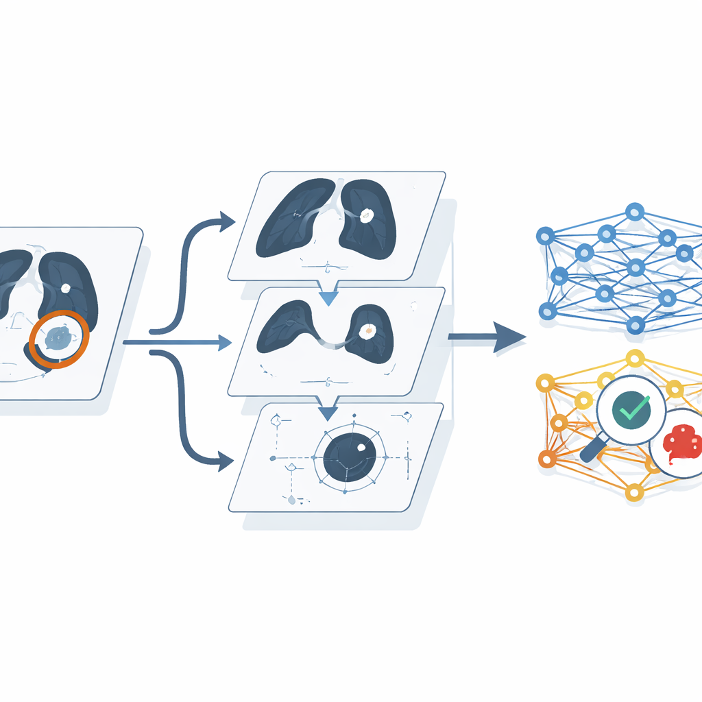
After cleaning the scans, the system automatically isolates the lung regions and then pinpoints individual lobes and nodules. It uses a segmentation model called Link-Net, which is designed to mark exactly which pixels in an image belong to lung tissue and to potential nodules. To help the computer learn robust patterns rather than memorizing a limited set of examples, the researchers augment the data: they rotate, flip, and partially erase sections of images in controlled ways, creating many realistic variations from each original scan. They also compute simple shape measures for each nodule, such as its area, perimeter, how irregular its outline is, and how compact it appears. These shape clues resemble what a radiologist informally judges when deciding whether a nodule seems suspicious.
How the new smart model reads the scans
The core of the work is a new artificial intelligence model called WISeRKNet, which combines two powerful deep-learning components. One part, based on a “wide slice residual” network, excels at examining whole CT slices and capturing subtle differences in lung texture and structure over relatively large regions. The other part, a “Kronecker” network, is designed to handle high-dimensional image patterns efficiently, allowing the system to learn complex relationships in the data without exploding in size. WISeRKNet fuses the information from the cleaned images and the shape measurements, then passes this richer description of each nodule through both network branches before making a final judgement about whether lung cancer is present.
Putting the model to the test
The researchers tested WISeRKNet on two publicly available collections of lung CT images. These datasets include scans of patients with malignant tumors, benign nodules, and healthy lungs, annotated by experts. They varied how much data the model could learn from, used standard cross-validation to avoid overestimating performance, and compared their approach with several existing computer methods, including traditional machine-learning models and other deep networks. Across a range of tests—such as overall accuracy, how often true cancers were correctly flagged, and how often healthy cases were correctly recognized—WISeRKNet consistently outperformed the alternative approaches. It maintained relatively strong results even when artificial noise was added to the images, suggesting that it is robust to less-than-perfect scans.
What this could mean for future care
In plain terms, the study shows that a carefully designed combination of cleaner imaging, smart use of shape information, and specialized deep-learning networks can detect lung cancer on CT scans with about nine correct decisions out of ten. While the authors note that the model is computationally demanding and not yet ready for real-time use in busy clinics, they plan to streamline the design so it can run faster and on more modest hardware. If successfully translated into practice, systems like WISeRKNet could become valuable assistants to radiologists, helping to spot dangerous nodules earlier and reducing missed cancers, ultimately improving outcomes for patients at risk.
Citation: Shanthi, A., Satheesh Kumar, S. & Koppu, S. WISeRKNet: wide slice residual Kronecker network for lung cancer detection based on CT images. Sci Rep 16, 9958 (2026). https://doi.org/10.1038/s41598-026-39793-w
Keywords: lung cancer, CT imaging, deep learning, computer-aided diagnosis, medical image analysis