Clear Sky Science · en
Non-invasive stroke volume assessment during cardiopulmonary exercise testing provides additional insight beyond O2-pulse in hypertrophic cardiomyopathy
Why this matters for people with heart muscle thickening
Many people with hypertrophic cardiomyopathy—a condition where the heart muscle is abnormally thick—struggle with shortness of breath and fatigue during everyday activities. Doctors usually assess how well these hearts cope with exercise using a breathing test, but this test only estimates how much blood the heart pumps with each beat. This study explored a simple, non-invasive way to directly track the heart’s pumping during exercise, uncovering hidden problems that standard tests can miss.
Looking more closely at the exercising heart
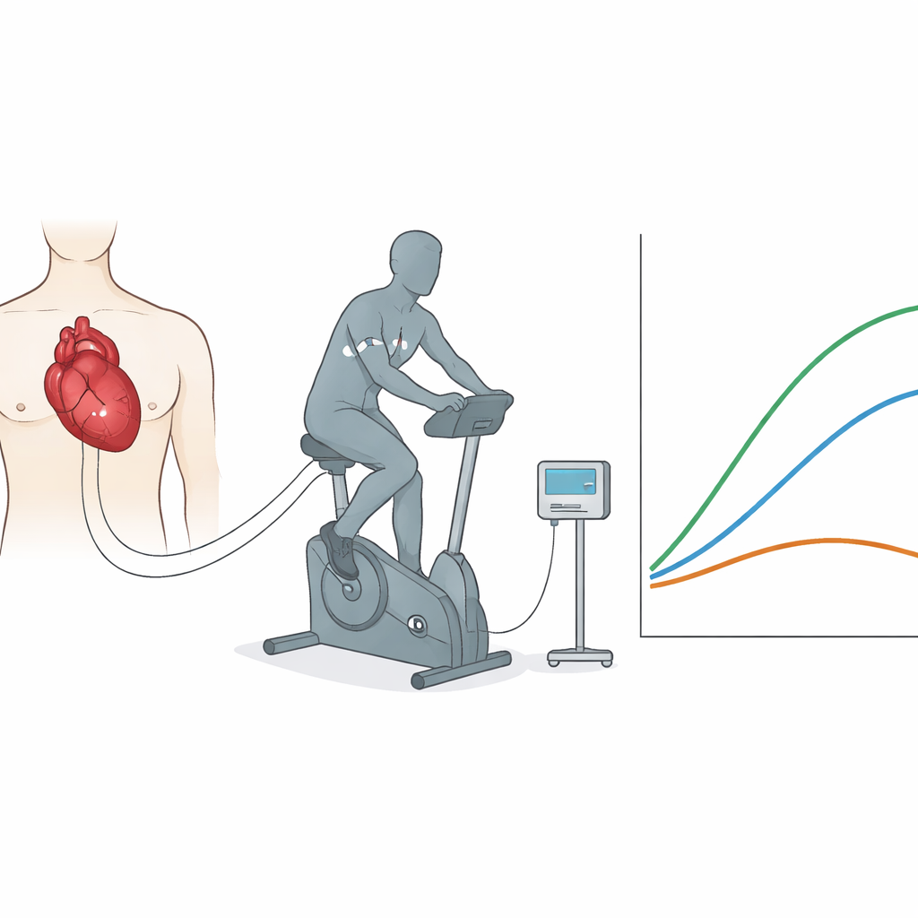
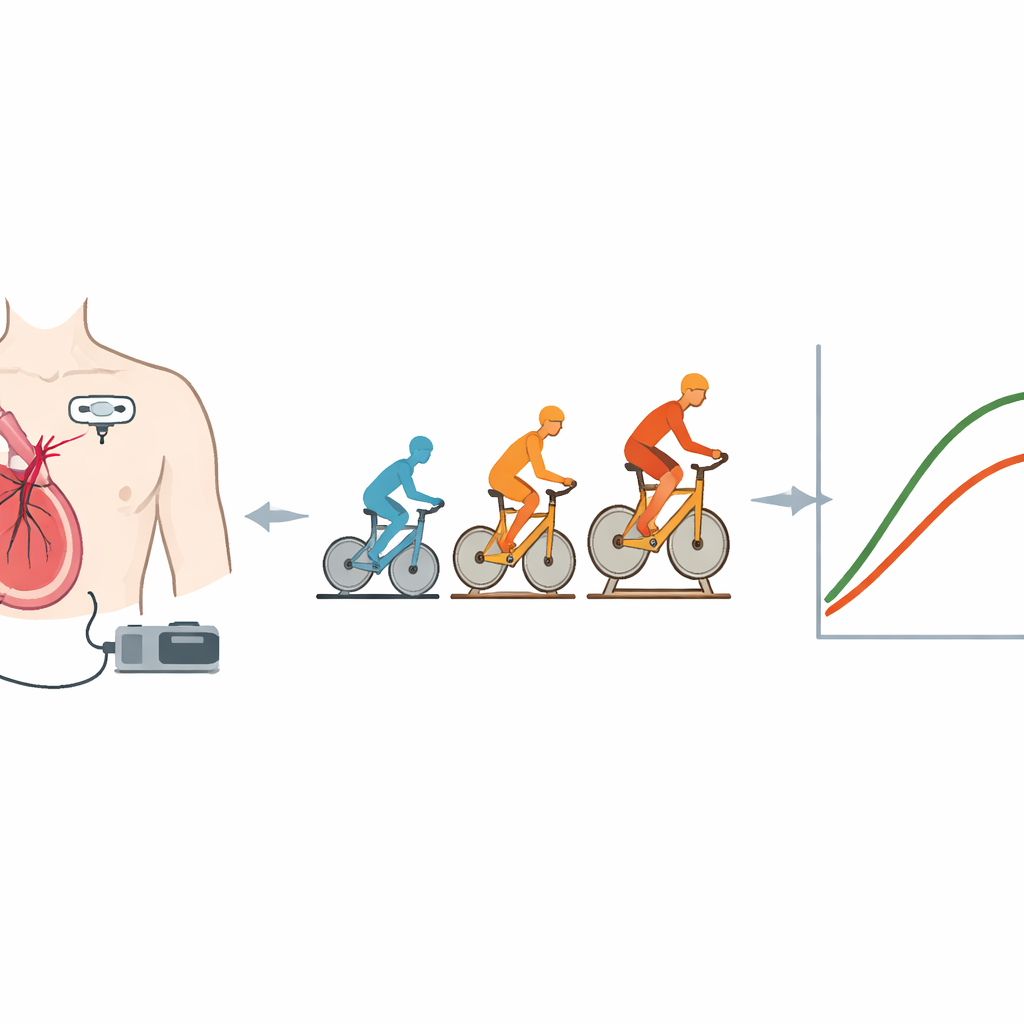
When we exercise, the heart must pump more blood with each beat, and we breathe faster to deliver more oxygen to the body. In clinics, a cardiopulmonary exercise test measures breathing and oxygen use while a person cycles on a stationary bike. A common shortcut in this test, called O2-pulse, uses breathing data and heart rate to roughly estimate how much blood is pumped with each heartbeat. But this shortcut can be fooled: muscles can sometimes extract more oxygen from a smaller amount of blood, hiding a fall in the true pumped volume. The researchers wanted to know whether directly tracking the heart’s stroke volume—how much blood leaves the heart with each beat—during exercise would give clearer insight in people with hypertrophic cardiomyopathy.
A new way to watch blood flow beat by beat
The team studied 102 adults with hypertrophic cardiomyopathy at two Italian specialty centers. Most had the non‑obstructive form of the disease, where the thickened heart muscle does not strongly block blood flow out of the heart at rest. All participants performed a maximal cycling test while breathing through a mouthpiece that measured oxygen and carbon dioxide. At the same time, a small device called PhysioFlow was connected via stickers on the chest to track changes in electrical impedance, a signal that can be used to estimate stroke volume and overall cardiac output beat by beat—without catheters, injections, or added discomfort.
Hidden pumping problems revealed
On paper, the group’s exercise capacity looked only mildly reduced, and average O2-pulse values were near normal. When doctors visually inspected the O2-pulse curve during the test, only 12% of patients seemed to have an abnormal pattern, such as flattening or falling values at higher workloads, which usually suggests the heart is failing to increase its output. However, the PhysioFlow data told a different story. Almost 40% of patients showed an abnormal stroke volume pattern: instead of continuing to rise, their stroke volume either plateaued early or decreased as the effort intensified. In every case where O2-pulse looked abnormal, stroke volume was indeed abnormal—but PhysioFlow uncovered more than twice as many patients with impaired pumping whose O2-pulse still looked normal.
What abnormal heart pumping means for breathing
Patients whose stroke volume behaved poorly during the final quarter of exercise had more trouble with gas exchange. They tended to breathe more for the same amount of oxygen used and carbon dioxide produced, and their end-tidal carbon dioxide—the level of carbon dioxide at the end of an exhaled breath—was lower. These patterns suggest that their hearts could not raise blood flow enough to match the muscles’ demands, pushing strain onto the lungs and blood vessels in the lungs. By contrast, patients whose stroke volume continued to rise in the last part of exercise reached a higher threshold before switching to predominantly anaerobic metabolism, converted exercise work into oxygen uptake more efficiently, and showed healthier breathing patterns.
Rethinking how we test and treat this condition
The study shows that relying on breathing measures alone can underestimate how many people with hypertrophic cardiomyopathy have real pumping limitations during exertion. A simple add‑on device that tracks stroke volume non‑invasively during standard exercise testing can reveal subtle problems in heart output that may explain symptoms and guide treatment decisions. In plain terms, even when usual test results look acceptable, many of these thickened hearts are quietly failing to boost the volume of blood they pump at higher effort, shifting burden to the lungs and limiting how far patients can push themselves. Integrating non‑invasive stroke volume monitoring into routine exercise testing could therefore give doctors a clearer picture of each patient’s functional reserve and help tailor care in an era of emerging targeted therapies.
Citation: Mapelli, M., Baracchini, N., Campana, N. et al. Non-invasive stroke volume assessment during cardiopulmonary exercise testing provides additional insight beyond O2-pulse in hypertrophic cardiomyopathy. Sci Rep 16, 9465 (2026). https://doi.org/10.1038/s41598-026-39769-w
Keywords: hypertrophic cardiomyopathy, exercise testing, stroke volume, noninvasive monitoring, cardiac output