Clear Sky Science · en
Age-dependent development and microarchitecture of the osteochondral unit of the humeral head in harbour porpoises (Phocoena phocoena)
How Porpoise Shoulders Grow Strong in the Sea
The way a young animal’s joints grow helps determine how well it can move for the rest of its life. For harbour porpoises, small toothed whales that dart through cold coastal waters, their front limbs act as stiff flippers that steer and stabilize them. This study looks inside the shoulder joint of harbour porpoises at different ages to see how the smooth, load‑bearing surface where bone and cartilage meet matures in an underwater world, and how that process compares with what we know from land mammals like horses, pigs, and rabbits. 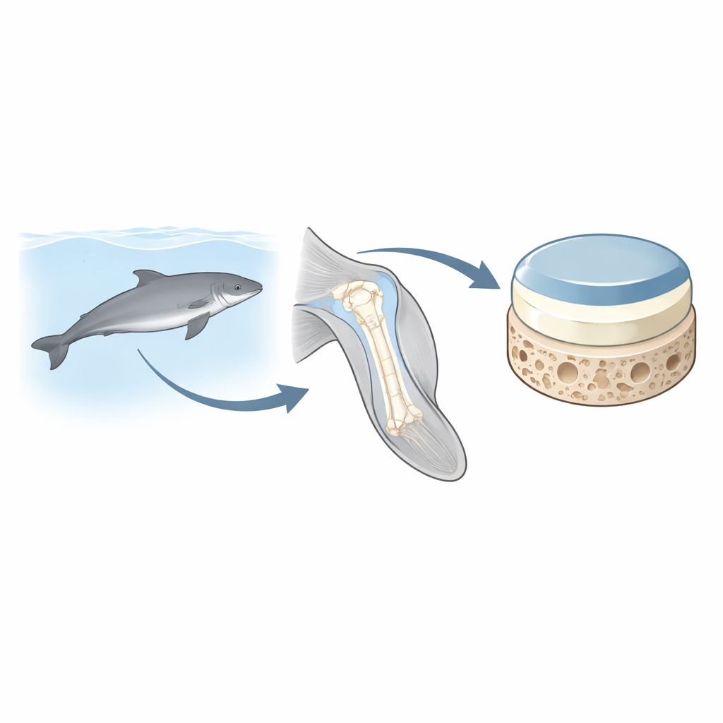
Bones and Joints Built for Life in Water
Whales, dolphins, and porpoises descended from land‑dwelling ancestors, but their skeletons have been reshaped for life in water. Hind limbs have mostly vanished, while the forelimbs became short, broad flippers. In harbour porpoises, most joints within the flipper are stiff or fused, yet the shoulder joint where the upper arm bone (the humerus) meets the rest of the body still moves. That joint is lined with a composite system of smooth cartilage sitting on bone, called the osteochondral unit. On land, we know that this structure changes rapidly after birth as young animals begin to stand, walk, and run. In contrast, the way this joint surface develops in fully aquatic mammals—with buoyancy, drag, and very different forces—has remained largely unknown.
Comparing Young and Adult Porpoise Shoulders
The researchers examined the rounded head of the humerus from seventeen harbour porpoises that had died naturally or after stranding. They grouped the animals as neonates, juveniles, and adults based on body length and sexual maturity, and then measured the size and shape of the flippers and humeral heads. Thin slices from the central, most heavily loaded part of the joint were stained and studied under the microscope, including with polarized light to reveal how the tough collagen fibers are arranged. The team also measured basic chemical components of the cartilage: DNA (a proxy for cell density), glycosaminoglycans that help the tissue hold water, and collagen, the main structural protein.
Slowly Shaping the Cartilage–Bone Interface
In newborn porpoises, the joint surface was covered by a relatively thick blanket of cartilage that had not yet separated into distinct zones. The topmost layer already showed fibers lying parallel to the surface, but deeper regions were more uniform and filled with rounded cells, plus blood‑vessel channels typical of growing cartilage. Notably, two key features seen in adult land mammals—a calcified cartilage band and a dense subchondral bone plate directly beneath the joint surface—were entirely absent. Juveniles began to show clearer layering in the cartilage and early hints of calcification near the cartilage–bone boundary, especially in larger youngsters, yet a true bone plate had still not formed. Only in adults did the researchers find a fully stratified structure with four recognizable cartilage layers, an irregular but continuous calcified zone, and a well‑developed bone plate anchoring the surface. At the same time, cell density fell with age, while cartilage matrix components increased, mirroring patterns seen in terrestrial animals. 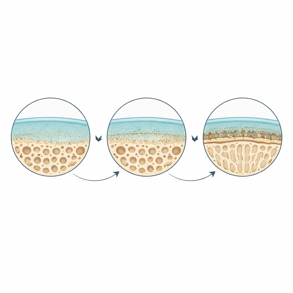
Collagen Arches That Arrive Late
A striking difference from land mammals lay in the timing and appearance of the collagen network that reinforces the cartilage. In many species that walk or run, the characteristic arch‑like pattern of fibers—often called Benninghoff arcs—emerges relatively early in life, within weeks or months. In porpoises, however, the collagen in deeper layers remained mostly oblique and disorganized through the juvenile stage. Only in adults did the classic arrangement appear, with an upper zone of fibers parallel to the surface, a middle zone of mixed directions, and a deep zone where fibers stand nearly perpendicular like pillars connecting cartilage to bone. The calcified layer and bone plate beneath also looked more wavy and irregular than in land animals, likely reflecting the gentler, differently directed forces on a joint that moves in water rather than bearing full body weight on land.
What This Means for Health, Evolution, and Repair
To a non‑specialist, the message of this work is that porpoise shoulder joints follow the same basic rules of growth as those of horses or sheep, but on a slower schedule and with shapes tuned to swimming rather than standing. The firm, layered interface between cartilage and bone still forms, and the collagen network still organizes into supportive arches, yet these milestones arrive only in adulthood and take on a more undulating form. These insights help explain how joints adapt to very different mechanical environments across evolution. They also provide a natural blueprint for engineers and doctors trying to design replacement tissues: if we want to build durable joint implants or repair damaged cartilage, we need to consider not only age and species, but also the specific loading environment—whether a limb is meant to push against the ground or slice through water.
Citation: Księżarczyk, M.M., IJsseldijk, L.L., van Weeren, P.R. et al. Age-dependent development and microarchitecture of the osteochondral unit of the humeral head in harbour porpoises (Phocoena phocoena). Sci Rep 16, 8466 (2026). https://doi.org/10.1038/s41598-026-39726-7
Keywords: harbour porpoise joints, articular cartilage development, aquatic mammal skeleton, osteochondral unit, mechanical loading and growth