Clear Sky Science · en
Resolving the contradiction between simulation and experimental results of using gold nanoparticles in proton therapy
Why tiny gold particles matter for cancer treatment
Proton therapy is a cutting-edge form of radiation treatment that can precisely target tumors while sparing surrounding healthy tissue. In recent years, scientists have tried mixing proton therapy with tiny specks of gold, called gold nanoparticles, to make the treatment even more lethal to cancer cells. Experiments show that this combo often kills more tumor cells than protons alone—but computer simulations have struggled to explain why. This paper tackles that long-standing mystery and points to a different main actor than many researchers expected.
Old story: blaming fast electrons
Gold nanoparticles are already famous in X‑ray and gamma‑ray treatments, where they boost damage mainly by spitting out swarms of energetic electrons. Those electrons travel short distances and break DNA in nearby cells. For years, many assumed the same basic story held for proton therapy: protons hit the gold, extra electrons fly out, and the cancer cells suffer. But there was a problem. Detailed computer models that track every particle and its energy—the kind used in this study—kept predicting very little extra dose in the cell nucleus from these electrons, especially because most nanoparticles sit in the cell’s outer region, not inside the nucleus where DNA lives. At the same time, lab experiments with cells showed clear increases in cell death and treatment effectiveness when gold was present. The numbers simply didn’t match.
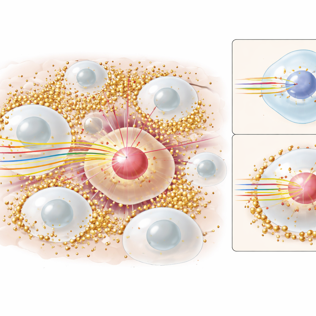
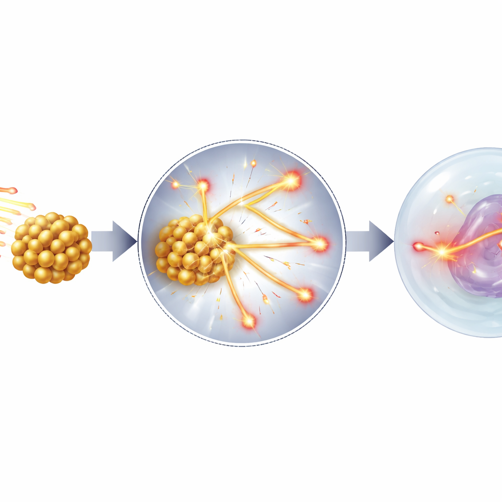
New picture: slowing the protons themselves
This work proposes and tests a different main mechanism: instead of acting mainly as electron emitters, gold nanoparticles behave as tiny speed bumps for protons. When a proton passes through a region dotted with high‑density, high‑atomic‑number metals like gold or iron, it undergoes many small collisions with those heavy atoms. Each collision drains a little more energy than it would in normal tissue, so the proton slows down more quickly and its energy loss per unit distance—known in physics as linear energy transfer, or LET—goes up. High‑LET tracks are especially damaging to DNA because they create dense clusters of breaks that the cell struggles to repair. By running detailed Monte Carlo simulations with the Geant4 toolkit, the author shows that gold and other heavy nanoparticles significantly boost the number of slow, high‑LET protons that reach the cell nucleus, even though the total path length is on the scale of micrometers, far beyond the reach of the low‑energy electrons traditionally blamed.
Matching simulations to real cell experiments
To see if this new picture holds up, the study recreates several published cell experiments where tumors were treated with proton beams plus various nanoparticles (gold, iron, and platinum) of different sizes and concentrations. For each case, the simulations compute how much extra dose the nucleus receives—summarized as a dose enhancement ratio—and then plug that into a standard radiobiology formula that relates delivered dose to cell survival. This approach modifies the usual curve that describes how many cells live or die after a given radiation dose. For most of the experiments examined, the predicted survival curves with nanoparticles lined up closely with the measured data, often within about one percent error. At the same time, the simulations show that the electron dose in the nucleus barely changes when nanoparticles are added, while the fluence of slower, more damaging protons clearly rises. A few mismatches remain, which the author attributes to uncertainties in how some experiments were set up or reported, but the overall trend strongly supports the proton-slowing explanation.
Limits, exceptions, and when gold helps most
The paper also explores situations where nanoparticles do not seem to help much. For very low‑energy proton beams that stop within just a few cell layers, there simply is not enough distance for the protons to encounter many nanoparticles and slow down meaningfully, so no strong boost in effectiveness is seen. Likewise, some complex nanoparticle shapes or poorly described experimental geometries are hard to reproduce in simulations, which may explain a few outliers where models and measurements disagree. The author notes that if ultra‑small particles actually enter the nucleus, then electron emission and chemical reactions with cell molecules could add to the effect. Still, across many realistic treatment conditions, the dominant pattern is consistent: more slowing of protons in gold‑rich regions leads to more concentrated damage in the nucleus.
What this means for future cancer care
For non‑specialists, the take‑home message is that gold nanoparticles in proton therapy work less like tiny electron guns and more like invisible brakes that turn fast, relatively gentle protons into slower, heavier hitters right where it matters most—the tumor cell’s DNA. By clarifying this mechanism and showing that it can accurately reproduce real cell survival data, the study helps resolve a long‑running conflict between theory and experiment. This insight could guide smarter designs of nanoparticle‑based treatments, such as choosing materials, sizes, and concentrations that maximize proton slowing near tumor nuclei while minimizing side effects. In the long run, this could make proton therapy more precise and powerful, offering better outcomes for patients with hard‑to‑treat cancers.
Citation: Tabbakh, F. Resolving the contradiction between simulation and experimental results of using gold nanoparticles in proton therapy. Sci Rep 16, 8012 (2026). https://doi.org/10.1038/s41598-026-39621-1
Keywords: proton therapy, gold nanoparticles, radiosensitization, cancer radiotherapy, nanomedicine