Clear Sky Science · en
Electron and focused ion beam microscopy of fossilized Albertosaurus sarcophagus (Dinosauria: Theropoda) bone reveals nano to microscale features
Peering Inside Dinosaur Bones
For anyone who has ever stared at a dinosaur skeleton in a museum and wondered what lies beneath the surface, this study offers a rare, ultra-close look. Researchers used advanced microscopes to zoom from the visible cross-section of an Albertosaurus leg bone all the way down to structures thousands of times thinner than a human hair. Their work shows that the internal architecture of bone, and even traces of its original building blocks, can survive for more than 70 million years.
Why Tiny Bone Details Matter
Bone is not a simple rock-like material. In living animals, it is a sophisticated composite built from tough protein fibers and hard mineral crystals, arranged in a precise hierarchy from whole limbs down to nanometer-scale patterns. When an animal dies and its bones fossilize, groundwater and buried sediments alter this delicate structure, replacing some parts with new minerals and changing others. By examining a thin slice of a juvenile Albertosaurus fibula (a slender lower-leg bone), the authors set out to see how much of that original architecture remains, and what the patterns of new minerals can tell us about the animal’s life and burial environment.
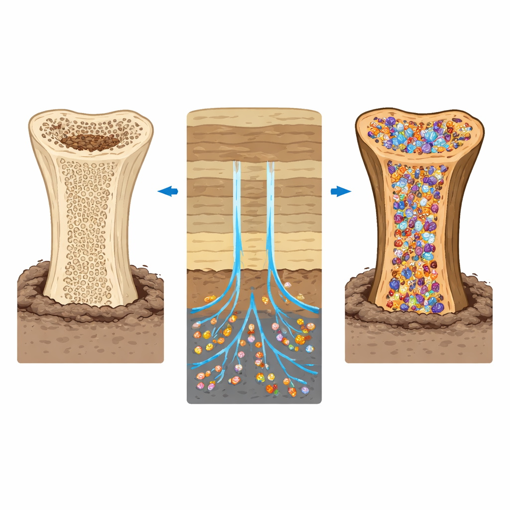
Minerals Flow In After Death
Using electron microscopes paired with chemical mapping tools, the team first explored how new minerals had invaded the fossil bone. They found that the original bone mineral, a form of calcium phosphate, was still present but now accompanied by a rich assortment of newcomers, including calcite, quartz, clay minerals, barium sulfate, and iron sulfide (pyrite). These materials had seeped in through the bone’s natural pore system—the central canals that once carried blood, the fine channels that connected bone cells, and even cracks formed during burial. In many places, the canals were lined or completely filled with these secondary minerals, recording pulses of groundwater movement and chemical change long after the dinosaur died.
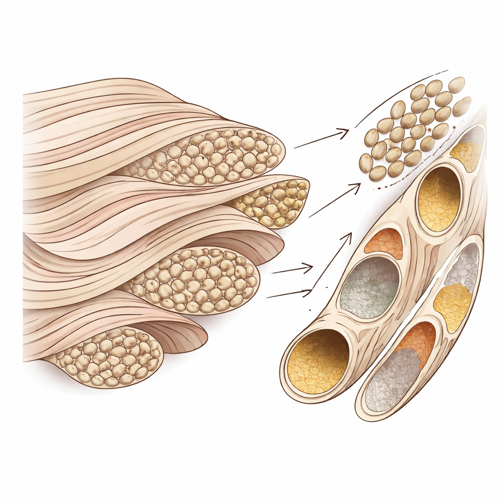
Ghosts of Cells and Fibers
At a finer scale, the researchers examined the tiny cavities that once housed bone cells. Some of these spaces were partly or fully filled with dense crystal growths, echoing a process seen in very old human bone where dying cells become entombed by mineral. Elsewhere, cavities were empty enough for the microscopes to reveal delicate networks of fibers lining their walls. Three-dimensional imaging showed that these fibers, which form the skeleton of bone tissue, were still arranged in a loose web around the cell spaces and along narrow channels. Measurements of their repeating band pattern matched that of collagen, the main structural protein in modern bone, indicating that the original fiber architecture has been astonishingly well preserved.
Hidden Order in Growing Bone
Zooming out slightly, the team reconstructed how bundles of these fibers were organized across small regions of the bone. In some areas, the fibers ran mostly in one direction, a pattern associated with rapidly formed bone that supports fast growth. In other zones near blood canals, the fibers rotated gradually from layer to layer, creating a plywood-like texture linked to stronger, more mature tissue. This mix of patterns matches what is seen in fast-growing young animals today and supports earlier work showing that juvenile tyrannosaur relatives grew quickly, remodeling their bones as they aged.
Ancient Mineral Clusters That Mirror Modern Bone
One of the most striking findings came from mapping how mineral is grouped within the fiber network. Within the aligned fiber regions, the researchers identified hundreds of small, three-dimensional clusters of mineral shaped like elongated ellipsoids. These clusters lined up with the surrounding fibers and resembled “tessellated” mineral units recently discovered in human and other mammal bones. Although the fossil clusters were somewhat larger—possibly due to species differences or slow crystal growth during fossilization—their overall shape and arrangement suggest that the basic rules for how bone mineral spreads through the collagen framework have changed little since the age of dinosaurs.
What This Means for Dinosaur Bones
In simple terms, this study shows that dinosaur bones preserve far more than just their outer shape. Even after tens of millions of years, the inner scaffolding of fibers and mineral, and the pathways once used by cells and blood, can remain readable under the right microscopes. The Albertosaurus fibula still carries a record of how its bone was built during rapid juvenile growth, how fluids later percolated through it underground, and how mineral clusters assembled at the nanoscale much like they do in our own skeletons today. By combining high-resolution imaging with careful chemical analysis, the work links fossil bone directly to living bone, revealing a deep continuity in how vertebrate skeletons are constructed and how they endure through geological time.
Citation: Williams, A., Schumann, D., Mallon, J.C. et al. Electron and focused ion beam microscopy of fossilized Albertosaurus sarcophagus (Dinosauria: Theropoda) bone reveals nano to microscale features. Sci Rep 16, 8521 (2026). https://doi.org/10.1038/s41598-026-39588-z
Keywords: dinosaur bone structure, fossilization, electron microscopy, collagen preservation, biomineralization