Clear Sky Science · en
Structural brain analysis in focal upper limb dystonia
Why this matters for everyday health
Many people live with a little-known movement problem called focal upper limb dystonia, where the hand or arm twists or cramps in ways they cannot control. Musicians, writers, and others who depend on precise hand movements can suddenly find everyday tasks difficult or painful. This study asks a simple but important question: does this disabling condition come from visible damage or reshaping of the brain, or is the wiring mostly intact and the problem lies in how the system works together?
Looking for clues inside the brain
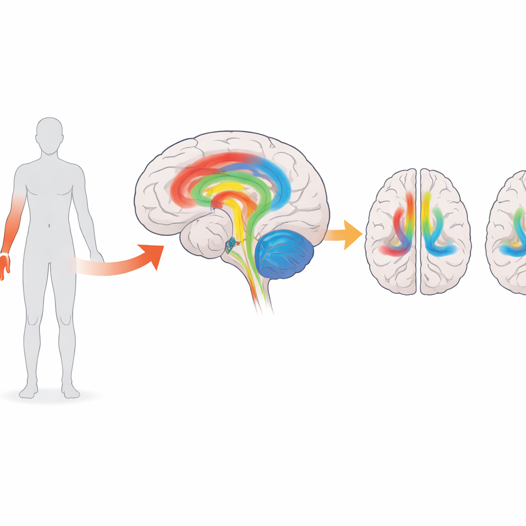
The researchers focused on a specific and relatively uniform group: 28 right-handed adults with dystonia affecting the right arm or hand, and 29 healthy people without movement problems. Everyone underwent detailed brain scans in a powerful 3‑Tesla MRI machine. The team collected two kinds of images: high‑resolution structural pictures that show the shape and thickness of the brain’s outer surface, and diffusion images that trace the pathways of the brain’s wiring, or white matter. By narrowing the study to one body region and one side, the scientists reduced the usual variation that can blur results in studies of more mixed patient groups.
Measuring brain shape and wiring
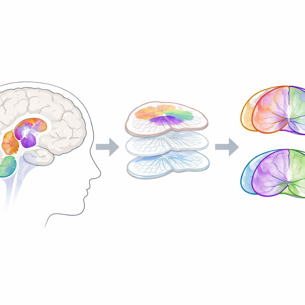
To examine the brain’s “grey matter,” where nerve cells live, the team used a method that carefully maps the thickness, area, and volume of the cortex instead of relying on older techniques prone to false alarms. They also measured key deep regions involved in movement, including the basal ganglia, thalamus, and cerebellum. For the “white matter” wiring, they followed major communication highways such as the corticospinal tract, fibers connecting the thalamus to the cortex, and bundles linking the two sides of the brain. Advanced statistical methods tested for even subtle differences, while taking into account age, sex, and head size.
What the scans did—and did not—show
Across all these measures, the result was striking in its consistency: the brains of people with focal upper limb dystonia looked structurally normal compared with healthy volunteers. The outer surface of the brain showed no reliable differences in thickness or size. Deep structures that are often suspected in movement disorders did not differ in volume. The brain’s wiring pathways, probed by diffusion imaging and tractography, also showed no clear changes in tissue quality. Even when the researchers asked whether people with more severe or longer‑lasting symptoms had different scan results, they found no meaningful patterns.
Rethinking the roots of dystonia
These findings fit with a growing view that dystonia is less about visible scars or shrinkage in the brain, and more about how networks of regions communicate in real time. Other work suggests that the timing and balance of signals among movement and sensory areas are disrupted, even when the underlying tissue looks intact. The preserved structure seen here may actually be good news: if the “hardware” of the brain is largely sound, treatments that adjust the “software”—such as deep brain stimulation or other forms of neuromodulation—may have more room to restore normal function. The authors argue that future studies using even finer imaging and combining structural and functional approaches, in larger groups, will be key to fully understanding and treating this puzzling but very real disorder.
Take‑home message
For people with focal upper limb dystonia, this study suggests that their disabling hand and arm symptoms are not due to obvious damage or loss of brain tissue. Instead, their brains appear structurally preserved, pointing toward problems in how brain networks operate rather than how they are built. This shift in perspective supports therapies that aim to rebalance brain activity, offering hope that carefully targeted stimulation or other network‑based treatments can ease symptoms without needing to repair broken structures.
Citation: de Faria, D.D., Paulo, A.J.M., de Paiva, J.P.Q. et al. Structural brain analysis in focal upper limb dystonia. Sci Rep 16, 9112 (2026). https://doi.org/10.1038/s41598-026-39542-z
Keywords: dystonia, brain MRI, movement disorders, white matter, deep brain stimulation