Clear Sky Science · en
Time course of visual plasticity following adult-onset deafness
When Hearing Fades, Vision Steps In
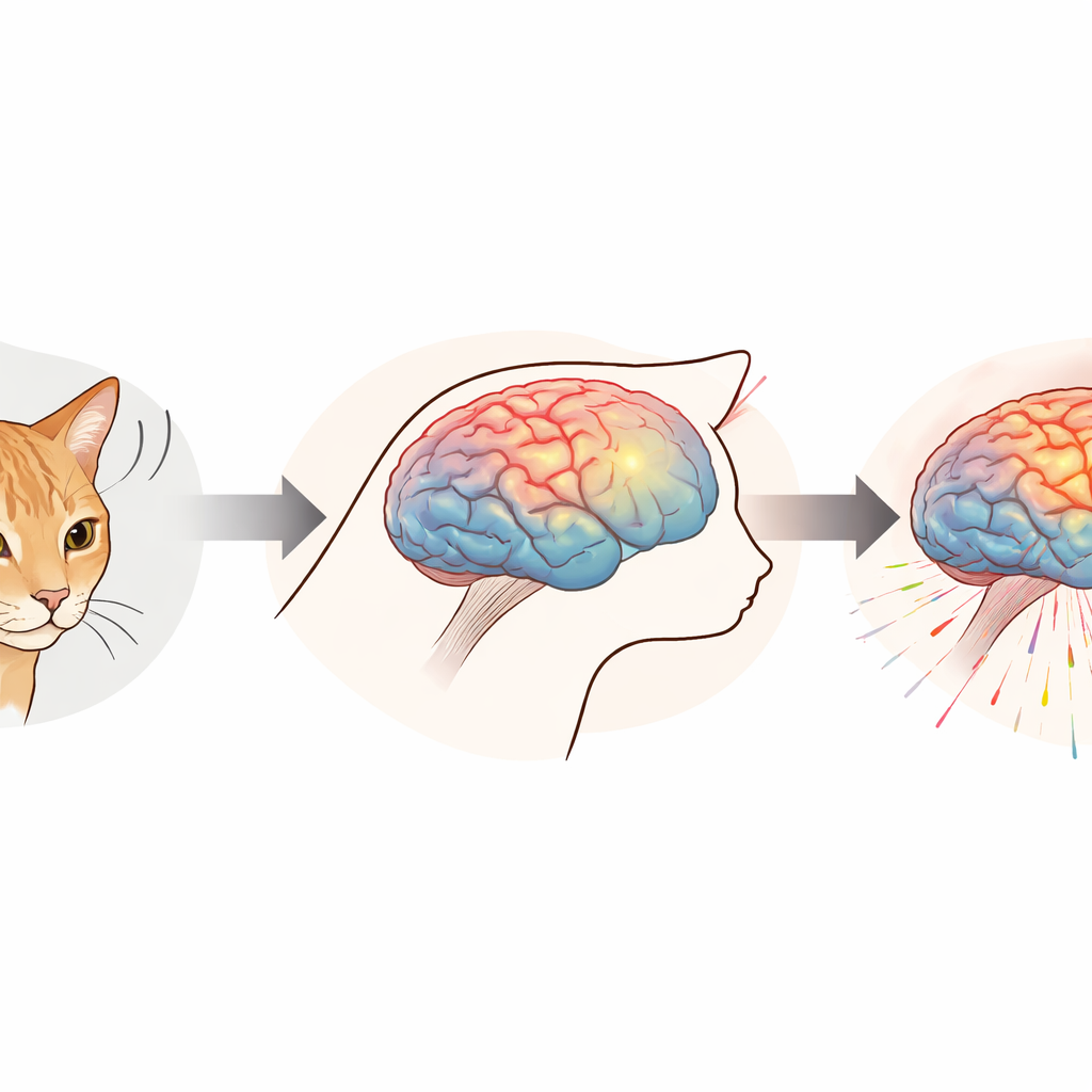
Many people lose their hearing well into adulthood, long after the brain’s early “critical periods” for development have passed. Doctors can restore some sound using devices like cochlear implants, but what happens in the silent months or years before treatment? This study uses an animal model to watch, in detail, how the brain’s visual responses change after hearing is lost in adulthood. The work offers a rare glimpse of how a mature brain reshapes itself and suggests new ways clinicians might track and perhaps harness this hidden plasticity.
Watching the Brain Adjust to Sudden Silence
The researchers studied four adult cats that were born with normal hearing and then intentionally made deaf using well-established medical procedures that damage the inner ear. Before and for more than a year after deafness, the team regularly recorded the animals’ brain responses to simple moving-dot patterns on a screen. These recordings, called visually evoked potentials, capture the tiny electrical signals produced when large groups of brain cells respond to a visual event. By placing small electrodes over regions roughly above visual and auditory brain areas, the scientists could follow how both "sight" and former "hearing" regions reacted to motion over time.
Signals That Grow Stronger and Faster
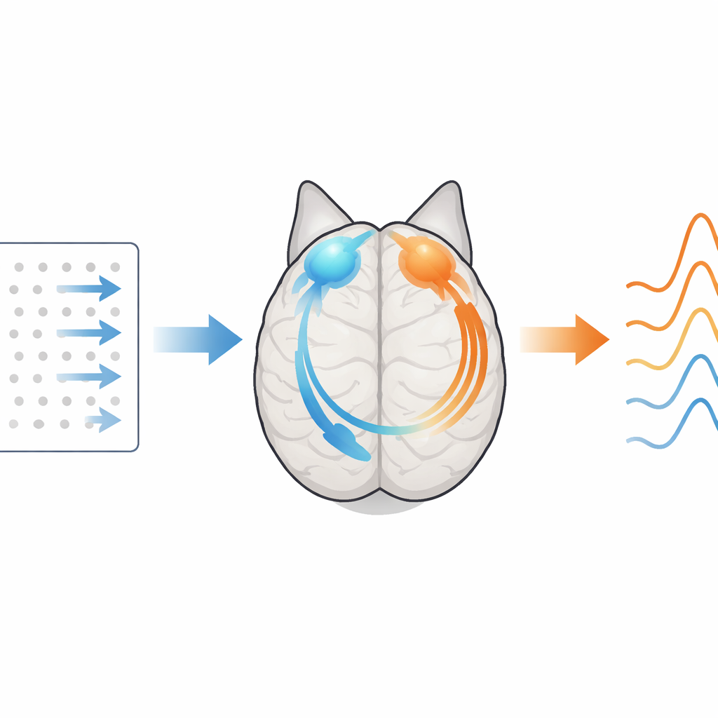
To track change, the team focused on two main features of the brain waves: overall signal power and the size and timing of a key positive bump in the waveform known as the P1 peak. After deafness, the visual responses recorded over the back of the head, where visual cortex sits, did not just stay stable—they grew. Within the first 100 days, the strength of these visual signals increased noticeably, and this amplification continued over the following months. The same pattern appeared, more slowly and more modestly, in the recordings over the temporal region, which normally houses auditory cortex. Here, visual signals strengthened only after roughly 200 days without hearing.
Different Brain Areas, Different Timelines
Timing changes added another layer to the story. As the months passed, the P1 peak arrived earlier, meaning the brain’s visual response became faster. Intriguingly, this speeding-up appeared sooner over the temporal site than over the visual site. In other words, the region that used to handle sound seemed to grow quicker at processing visual motion, even though its visual signals took longer to grow in size. This mismatch hints that different kinds of plasticity—boosting response strength versus speeding up processing—may unfold on distinct timelines in different parts of the brain.
How Motion Helps Reveal Hidden Changes
The visual test in this study was deliberately simple: a field of dots that suddenly began to move at different speeds. Earlier work in both deaf humans and animals has shown that motion detection is one of the visual skills that often improves after hearing loss. By using motion-onset stimuli, the researchers selected a probe that is both stable to measure and directly relevant to known behavioral gains. Their detailed analyses showed that the strongest differences between hearing and deaf states emerged when the dots moved at medium to high speeds, suggesting that brisk motion is especially sensitive to the brain’s rebalancing after deafness.
From Lab Recordings to Future Patient Care
Together, these findings show that even a fully grown brain can reorganize itself substantially after hearing loss: visual responses become stronger and faster not only in traditional visual areas but also in regions that once specialized in sound. Because visually evoked potentials are also widely used in human clinics, the same approach could help track how patients’ brains adapt during the silent interval before a cochlear implant is placed. In time, such measurements might guide when to intervene and how to tailor rehabilitation, turning an invisible phase of brain change into something doctors can see and potentially use to improve outcomes.
Citation: Zhu, S., Bao, X. & Lomber, S.G. Time course of visual plasticity following adult-onset deafness. Sci Rep 16, 9384 (2026). https://doi.org/10.1038/s41598-026-39490-8
Keywords: neuroplasticity, adult-onset deafness, visual evoked potentials, cross-modal reorganization, sensory compensation