Clear Sky Science · en
Establishing a robust preclinical model to investigate early and late radiation-induced skin reactions
Why this matters for people with cancer
Modern radiation therapy saves lives, but it can be tough on the skin, causing painful redness, peeling, and long‑lasting stiffness or scarring. Doctors know these reactions are linked to the radiation dose, yet it has been hard to study them in detail or to test new treatments safely. This paper describes a carefully designed mouse model that mimics both the early and late skin problems seen in patients, offering a practical test bed for gentler radiation schedules and protective therapies. 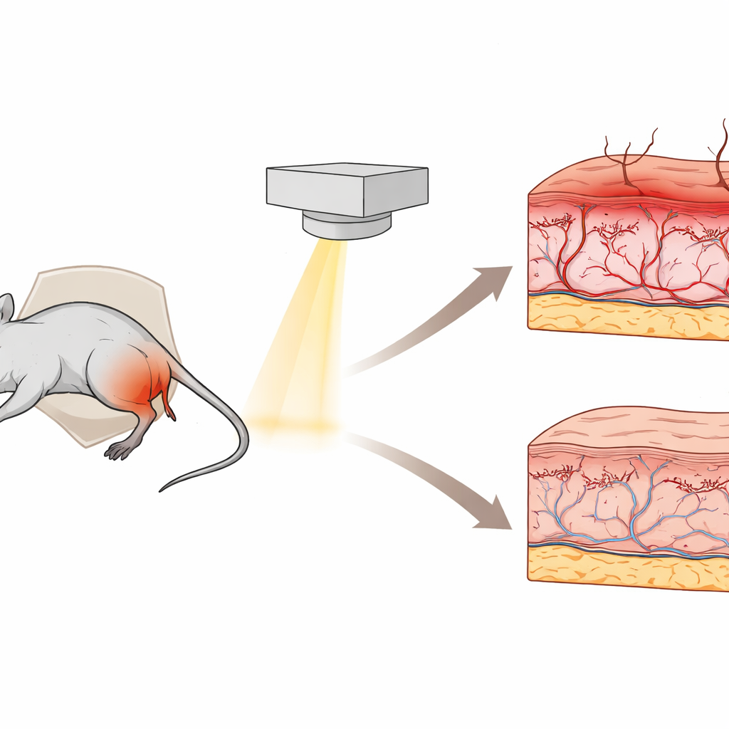
Turning a common side effect into a research focus
When a tumor is treated with radiation, nearby healthy skin often pays a price. Early changes can appear within days to weeks as redness, swelling, and peeling, while late changes—months or years later—can show up as hair loss, hardening, or scarring of the skin. These problems may delay treatment and lower quality of life, yet existing animal studies usually look only at short‑term damage or use single, very high radiation doses that do not resemble standard clinical care. The authors set out to build a more realistic model that tracks both early and late skin injury over time using fractionated radiation, the same basic approach used in human radiotherapy.
How the new mouse model was built
Researchers worked with male Swiss albino mice and focused radiation on a small area of the right hind limb, while shielding the rest of the body with lead. This setup allowed them to damage a well‑defined skin region without harming vital organs, similar to how a patient’s tumor is targeted while nearby tissues are protected. One group of mice received a total of 30 units of dose spread over three daily sessions; another group received 50 units over five sessions, better reflecting the repeated exposures used in the clinic. Over the next month, a dermatologist, unaware of which dose each mouse had received, scored visible skin changes using a standard clinical scale, and small skin samples were taken at days 15 and 30 for microscopic analysis.
What early skin reactions looked like
The visible scores showed a clear dose–response. The lower‑dose group typically developed moderate reactions that peaked around day 10 and healed by day 30. In contrast, the higher‑dose group developed more severe reactions, peaking a bit later and taking about five extra days to fully recover. Under the microscope, irradiated skin showed a thickened outer layer, greater numbers of inflammatory cells, and a sharp drop in hair follicles compared with unexposed skin on the other leg. The higher dose produced more intense inflammation, surface breakdown, and a special pattern of surface changes that signal rapid, stressed skin turnover. These features closely resemble what physicians see in patients with strong early skin reactions to radiation. 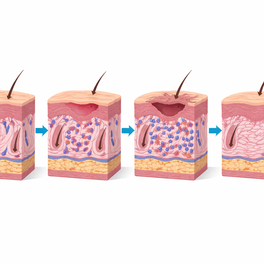
Tracing long‑term scarring and hair loss
To capture late effects, mice that had received the higher dose were followed for four months. Externally, the treated skin gradually lost hair and developed a firmer, somewhat indented feel, suggesting the onset of fibrosis—scar‑like thickening. Tissue stains that highlight collagen fibers confirmed this impression: the treated skin had thicker, more densely packed, and disorganized collagen in the deeper layer, together with a lasting loss of hair follicles. Scoring by a pathologist showed higher levels of inflammation, fibrosis, and cellular disruption in the treated skin than in untreated skin, while deeper muscle tissue showed milder changes. Together, these findings indicate that the same animals first developed an early, reversible reaction and later a more permanent, scar‑like state, echoing the course seen in human skin.
What this model means for future care
By recreating both short‑term irritation and long‑term scarring in a single, controlled mouse model that uses clinically relevant radiation schedules, this study offers a powerful tool for future work. Scientists can now probe how skin cells, immune cells, and supporting tissue respond over time, and they can systematically test drugs, dressings, or light‑based treatments designed to protect the skin or speed healing. Ultimately, insights gained from this model could help make radiation therapy not only effective against tumors but also kinder to the skin, reducing pain, avoiding treatment breaks, and improving quality of life for people undergoing cancer care.
Citation: Pai, P.A.N., Mumbrekar, K.D., Mahato, K.K. et al. Establishing a robust preclinical model to investigate early and late radiation-induced skin reactions. Sci Rep 16, 9064 (2026). https://doi.org/10.1038/s41598-026-39414-6
Keywords: radiation-induced skin reactions, radiotherapy side effects, skin fibrosis, preclinical mouse model, cancer treatment toxicity