Clear Sky Science · en
A field of view-based assessment of soft tissue calcifications in cone beam computed tomography of the maxillofacial region
Why tiny stones in the neck matter
Most people only think about dental X‑rays when they have a toothache or need braces. Yet modern 3D dental scans can quietly reveal far more than cavities. This study looked at small mineral deposits, or “little stones,” that can form in the soft tissues of the head and neck. Some of these deposits are harmless, but others may hint at problems such as blocked salivary glands or even an increased risk of stroke. Understanding how often these deposits appear, and where dentists are most likely to spot them, can turn a routine scan into an early‑warning system for wider health issues.
Looking beyond the teeth
When dentists order cone‑beam computed tomography (CBCT), they usually focus on a small region of the jaws to plan implants, find impacted teeth, or assess bone loss. But the scan also captures nearby soft tissues, including the tonsils, neck vessels, and ligaments. The researchers asked a practical question: if a scan is aimed mainly at the upper jaw (maxilla) or the lower jaw (mandible), how likely is it to show these hidden calcifications, and does this change with a patient’s age or sex? Answering this could help radiologists know where to look more carefully, and which patients might benefit from medical follow‑up.
How the scans were studied
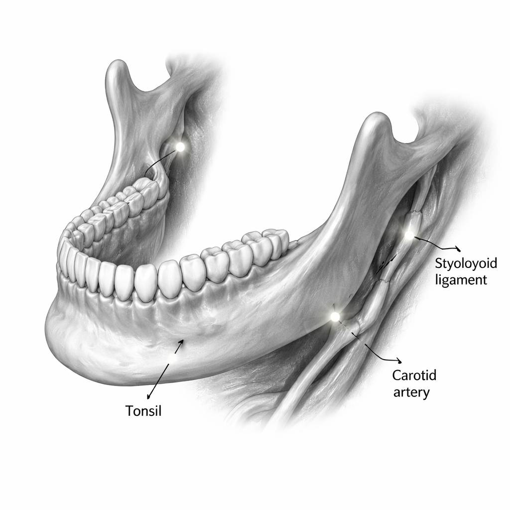
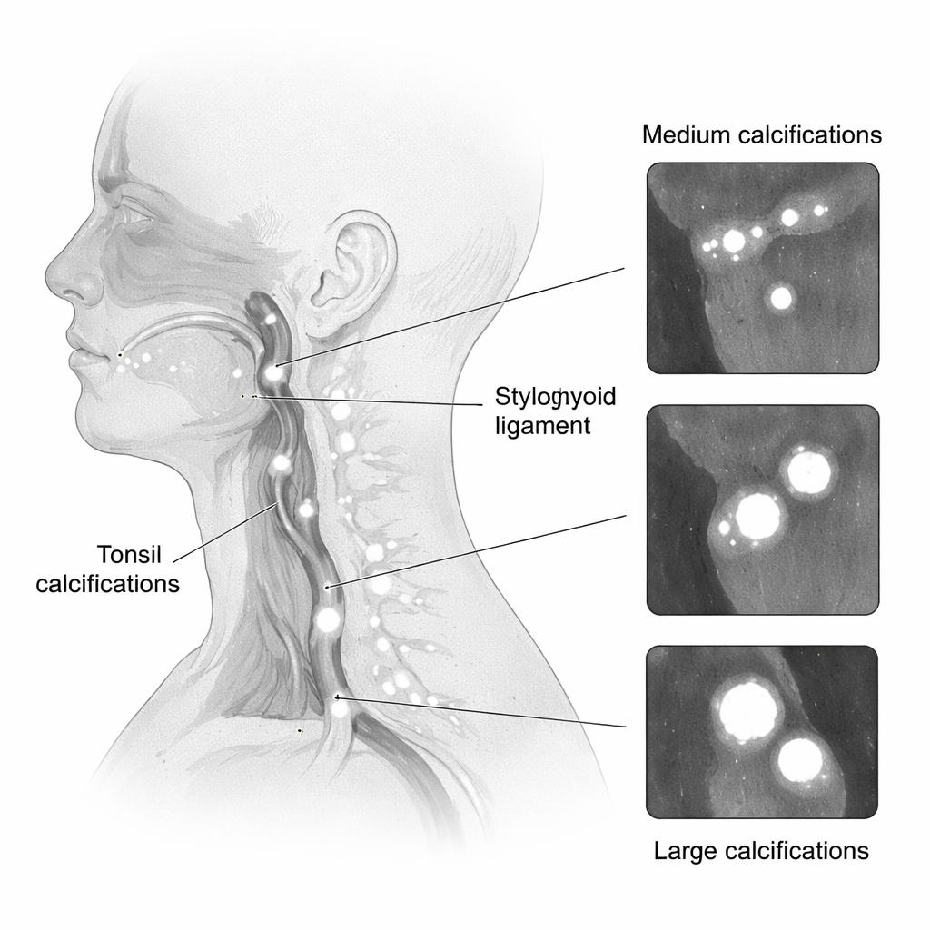
The team reviewed 420 CBCT scans taken between 2020 and 2024 at a university dental clinic in Iran. Each scan covered either the upper or lower jaw using a standardized field of view. Patients ranged in age from 6 to 82 years. Poor‑quality scans and those distorted by surgery or rare calcification disorders were excluded. Two experienced radiologists, trained on example cases beforehand, independently checked each scan for specific types of soft tissue calcification, such as tonsil stones (tonsilloliths), stiffened stylohyoid ligaments, salivary stones, calcified lymph nodes, and, in lower-jaw scans, calcified deposits in the carotid arteries of the neck. They measured the largest dimension of each finding and grouped them as small (1 millimeter or less), medium (1–3 millimeters), or large (3 millimeters or more). Agreement between the readers was almost perfect, meaning the findings were highly reliable.
What the researchers found
Soft tissue calcifications were common. Overall, they were about 1.3 times more likely to be seen in scans focused on the lower jaw than in those aimed at the upper jaw. Across both regions, tonsil stones were the most frequent type, followed by calcified stylohyoid ligaments. In lower‑jaw scans, about one in three patients had tonsil stones, and in upper‑jaw scans nearly one in five did. Most deposits, including tonsil stones and calcified ligaments, fell into the “medium” size range, large enough to be seen clearly on CBCT but often still unnoticed in everyday life.
Age, sex, and higher‑risk findings
Age turned out to be a strong predictor. As patients grew older, calcifications not only appeared more often but also tended to be larger. People over 50 had more than twice the odds of having tonsil stones and were far more likely to show calcified plaques in the carotid artery area on lower‑jaw scans. In upper‑jaw scans, large calcified ligaments were much more common after the early forties. There were also some sex‑related patterns: in the upper‑jaw field, women more often showed calcified stylohyoid ligaments, while men more often had skin‑related calcifications. Still, many calcification types were too rare for firm conclusions about sex differences. The authors emphasize that lower‑jaw scans, in particular, capture regions close to the throat and neck vessels, making them especially valuable for spotting tonsil stones and possible artery calcifications.
Turning incidental findings into early warnings
From a lay perspective, the main message is that dental 3D scans can quietly reveal more than dental problems alone. This study shows that small mineral build‑ups in the head and neck are common, especially as people age, and that lower‑jaw scans are more likely to catch them. While many deposits are harmless, some—such as those near the carotid arteries—may signal increased health risks and deserve medical attention. The authors propose a simple checklist for radiologists to follow whenever they read CBCT images, ensuring they consistently look for tonsil stones, artery calcifications, stiffened ligaments, and other soft tissue deposits. In older patients, especially those over 50, a careful review of these scans could support earlier referrals and potentially help prevent serious events, all without adding extra radiation or new tests.
Citation: Latifi Douabsari, M., Rahmanpanah, S., Khosravifard, N. et al. A field of view-based assessment of soft tissue calcifications in cone beam computed tomography of the maxillofacial region. Sci Rep 16, 7685 (2026). https://doi.org/10.1038/s41598-026-39388-5
Keywords: cone-beam CT, soft tissue calcification, tonsil stones, carotid artery calcification, dental imaging