Clear Sky Science · en
Radiomic profiling of chest CT in a cohort of sarcoidosis cases
Why lung scans hold hidden clues
Sarcoidosis is a disease that often attacks the lungs, leaving many people short of breath and unsure how their illness will progress. Doctors rely on chest scans to judge how badly the lungs are affected, but reading these images by eye can be subjective. This study asks a simple question with big implications: can computers measure subtle patterns in lung scans more precisely than the human eye, and do those patterns actually relate to how well people breathe and feel in daily life?
From pictures to numbers
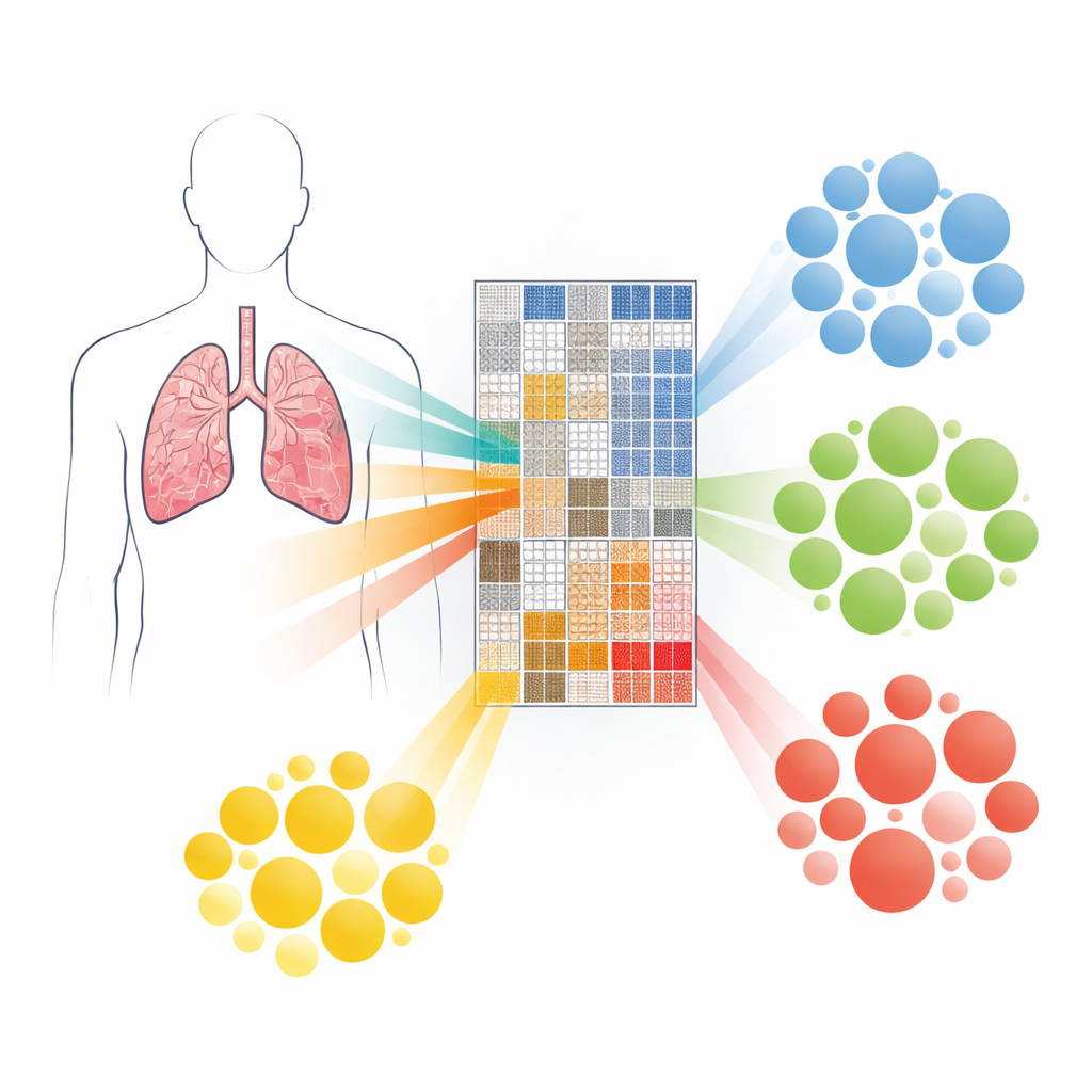
Traditionally, lung involvement in sarcoidosis is graded using chest X‑rays or high‑resolution CT scans that radiologists score by hand. These visual scores, while useful, can vary from one reader to another and may not fully capture the complexity of the disease. In this work, researchers turned to “radiomics,” an approach that converts every chest CT scan into hundreds of numerical features that describe how light and dark areas are distributed and how textures repeat across the image. Instead of focusing only on how dense the lung tissue appears, they also measured fine‑grained patterns in neighboring pixels, which are thought to reflect scarring and other microscopic changes.
Sorting patients into hidden groups
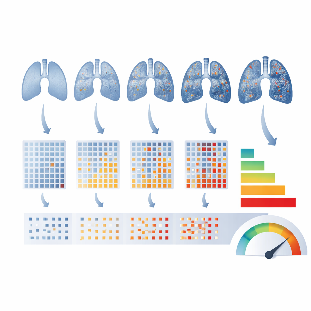
The team analyzed high‑resolution CT scans from 320 people with sarcoidosis who took part in a large U.S. research project. For each person, they calculated more than 500 radiomic features from both lungs and then used statistical methods to trim this down to a leaner set that avoided duplication. With these features, they applied a form of machine learning that looks for natural groupings in the data without being told ahead of time who is “mild” or “severe.” This unsupervised clustering uncovered four distinct radiomic profiles, each representing a different pattern of lung texture and density.
Linking scan patterns to lung strength
These four radiomic groups were then compared with standard clinical measures. Patients also had traditional X‑ray–based staging, detailed visual scoring of their CT scans, and lung function tests such as how much air they could blow out and how well oxygen moved from their lungs into the blood. The radiomic clusters did not simply mirror the usual staging system; each cluster contained a mix of classic stages. Yet the clusters were strongly tied to lung performance. People in the “healthiest” cluster had the best breathing test results, while those in the most abnormal cluster had much lower lung volumes and gas‑transfer ability. Overall, radiomic groupings explained about 10–15% of the variation in lung function beyond what could be accounted for by age, body size, and other basic traits.
What the textures reveal about daily life
The study also examined how these imaging patterns related to what patients reported about their symptoms, including fatigue, shortness of breath, and quality of life. The connections here were weaker than for the breathing tests but still telling. The most severely affected radiomic cluster not only showed more scarring and distortion on scans but was also linked with worse shortness of breath and poorer physical health scores. Another cluster was marked by obstructed airways and particularly low ratios on breathing tests, suggesting it represents a distinct “obstructive” form of disease. Together, these patterns hint that lung texture captured by radiomics can reflect meaningful differences in how sarcoidosis shows up in everyday experience, even if symptoms are influenced by many factors.
How this could change care
For people living with sarcoidosis, the promise of this work is a future in which a routine CT scan can be processed in minutes to yield an objective fingerprint of disease in the lungs. The findings suggest that radiomic profiles capture aspects of lung damage that traditional staging may miss and that these profiles are tied to how well the lungs function. While more research is needed—especially to track how these measures change over time—the study offers early evidence that computers reading textures in lung scans could help doctors classify disease more accurately, spot subtle changes sooner, and eventually tailor treatment to the specific pattern of lung injury each patient carries.
Citation: Carlson, N.E., Lippitt, W.L., Ryan, S.M. et al. Radiomic profiling of chest CT in a cohort of sarcoidosis cases. Sci Rep 16, 9695 (2026). https://doi.org/10.1038/s41598-026-39384-9
Keywords: sarcoidosis, radiomics, chest CT, lung function, medical imaging