Clear Sky Science · en
To explore the performance of ultrasound elastography in staging diabetic kidney disease: a systematic review and meta-analysis
Why kidney changes matter in diabetes
For millions of people living with type 2 diabetes, the silent damage it can inflict on the kidneys is one of the most serious long-term threats. By the time standard blood and urine tests clearly signal trouble, much of the harm may already be done. This study asks a simple but important question: can a newer, painless ultrasound technique that measures how “stiff” the kidney tissue is help doctors spot diabetic kidney disease earlier and track how it worsens over time?
A gentle scan that feels for stiffness
Ultrasound elastography is an add-on to ordinary ultrasound. Instead of just drawing a picture of the kidney, it sends tiny vibrations through the tissue and measures how fast they travel. Stiffer tissue – usually a sign of scarring and long-term injury – lets waves move faster. In this review, researchers gathered results from 18 previous studies including more than 2,700 people: some were healthy, some had diabetes without kidney damage, and others had various stages of diabetic kidney disease. Across all of these groups, they focused on one main number: cortical stiffness, a measure of how rigid the outer working layer of the kidney had become.
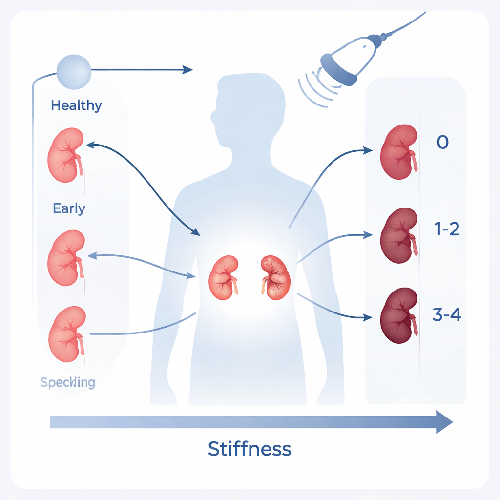
From healthy to harmed: a rising stiffness scale
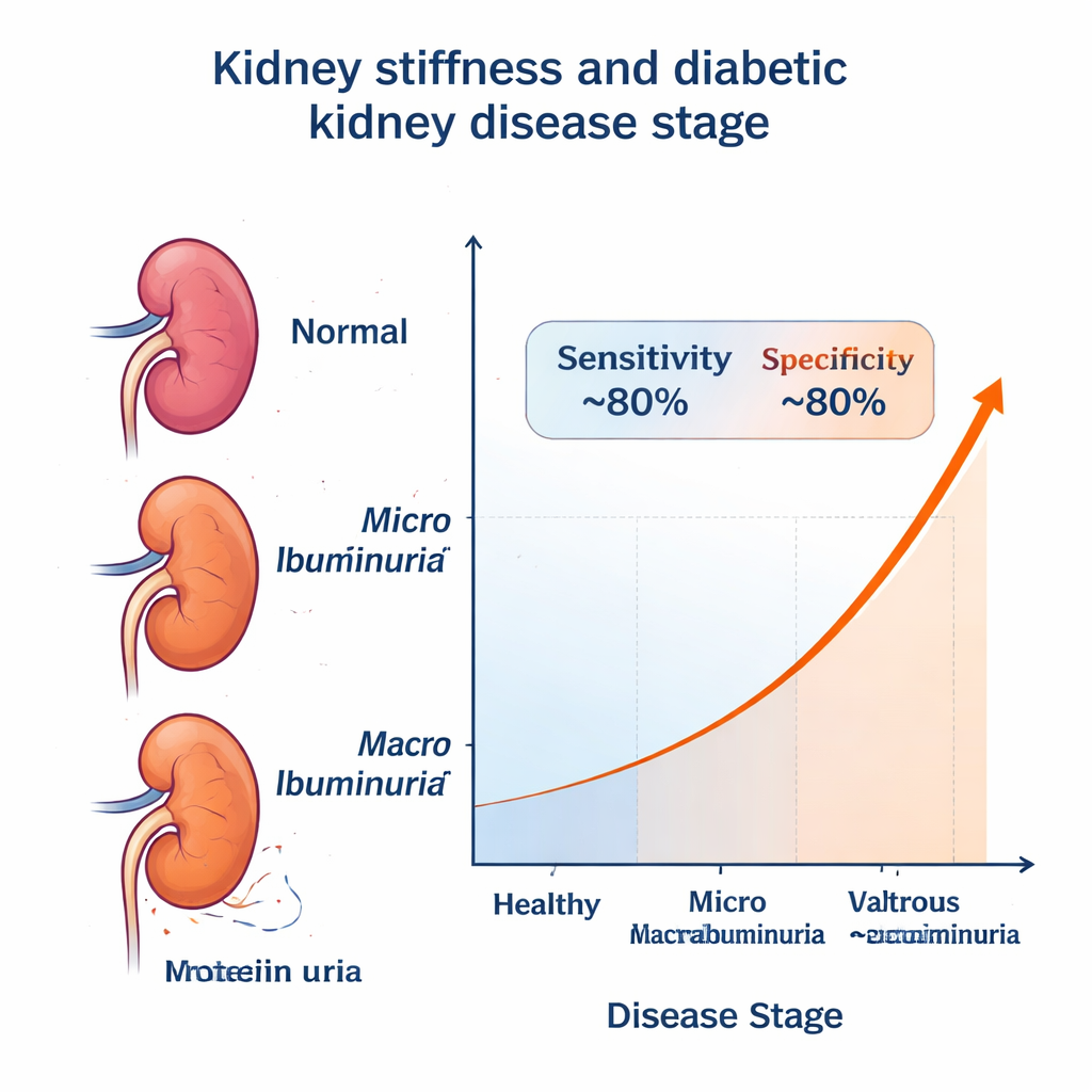
The team found a clear pattern. On average, healthy volunteers had the softest kidneys. People with diabetes but no kidney disease already showed stiffer cortices. Stiffness rose further in those with early signs of damage (tiny amounts of protein leaking into the urine, called microalbuminuria), and was highest in those with more advanced disease (larger protein leaks, or macroalbuminuria). In percentage terms, kidney stiffness was roughly one-fifth higher in people with diabetes but no diagnosed kidney disease than in healthy controls, and then increased again as kidney damage and urine protein levels worsened. These findings match what doctors know from biopsies: as diabetic kidney disease progresses, normal tissue is gradually replaced by scar-like material that is denser and less elastic.
How well can stiffness flag dangerous stages?
Beyond broad trends, the key test is whether elastography can tell one patient’s stage from another’s. For studies that reported enough detail, the authors grouped participants into simpler categories. First, they asked whether stiffness could separate people without diabetic kidney disease from those with any stage of it. Here, the scan correctly flagged disease about 79 percent of the time and correctly reassured about 83 percent of those without it. The overall diagnostic score, known as area under the curve, was 0.88 on a scale where 1.0 is perfect and 0.5 is no better than guessing. In a second analysis, they asked whether stiffness could distinguish milder stages from clearly advanced disease. The performance was similar: about 80 percent sensitivity and 79 percent specificity, with an area under the curve of 0.87.
Promise and practical limits
Although the average numbers march upward from healthy to severely diseased, the ranges overlap. Some patients with early disease have quite stiff kidneys, while a few with more advanced disease appear softer on the scan. That means elastography alone cannot perfectly label an individual’s stage. The authors argue it should be treated as a new piece of the puzzle to be read alongside standard blood tests, urine protein levels, and blood pressure, not as a stand‑alone gatekeeper. They also note that results can vary with the operator’s skill, the exact ultrasound machine used, and physical factors such as body size. Many of the original studies left out patients with other common illnesses like high blood pressure or liver disease, which may limit how well the findings reflect everyday clinic populations.
What this means for people with diabetes
For a person with type 2 diabetes, the main message is that their kidneys may begin to change long before standard tests clearly show damage – and that a simple, non-invasive scan of tissue stiffness could help bring those changes to light. This review concludes that ultrasound elastography is a promising, radiation‑free tool for staging diabetic kidney disease and spotting more serious forms, with good—but not perfect—accuracy. If future work standardizes how the scans are performed and confirms these results in broader patient groups, doctors may gain a new way to track kidney health over time and adjust treatment earlier, potentially slowing or preventing kidney failure.
Citation: Mohebbi, A., Mohammadzadeh, S., Asli, F. et al. To explore the performance of ultrasound elastography in staging diabetic kidney disease: a systematic review and meta-analysis. Sci Rep 16, 7542 (2026). https://doi.org/10.1038/s41598-026-39278-w
Keywords: diabetic kidney disease, ultrasound elastography, kidney stiffness, type 2 diabetes, noninvasive imaging