Clear Sky Science · en
Synaptic and cytoskeletal CSF signatures of motor neuron disease: the role of cyclase-associated protein 2
Why Nerve Connections Matter in Movement Disease
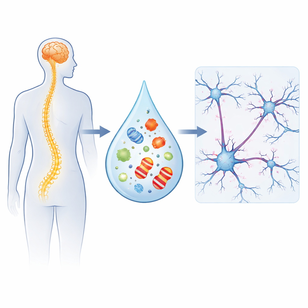
Motor neuron disease, which includes amyotrophic lateral sclerosis (ALS), robs people of their ability to move, speak, and eventually breathe. While we know that the nerve cells controlling muscles die, scientists are increasingly realizing that the earliest trouble may start at the tiny contact points where nerve cells talk to each other and to muscles. This study asks a simple but powerful question: can we detect early changes at these connections by looking at proteins floating in the fluid that bathes the brain and spinal cord?
A Closer Look at Brain and Spinal Fluid
The researchers focused on the clear liquid called cerebrospinal fluid, which surrounds the brain and spinal cord and can mirror what is happening inside them. They compared samples from 60 people with motor neuron disease and 40 healthy volunteers of similar age. In these samples, they measured several known markers of nerve injury and support cells, along with two proteins tied to the communication points between nerve cells: SNAP-25, found at the sending side of the connection, and CAP2, found mainly at the receiving side and closely linked to the internal scaffolding of the cell.
A New Signal from the Receiving Side of Synapses
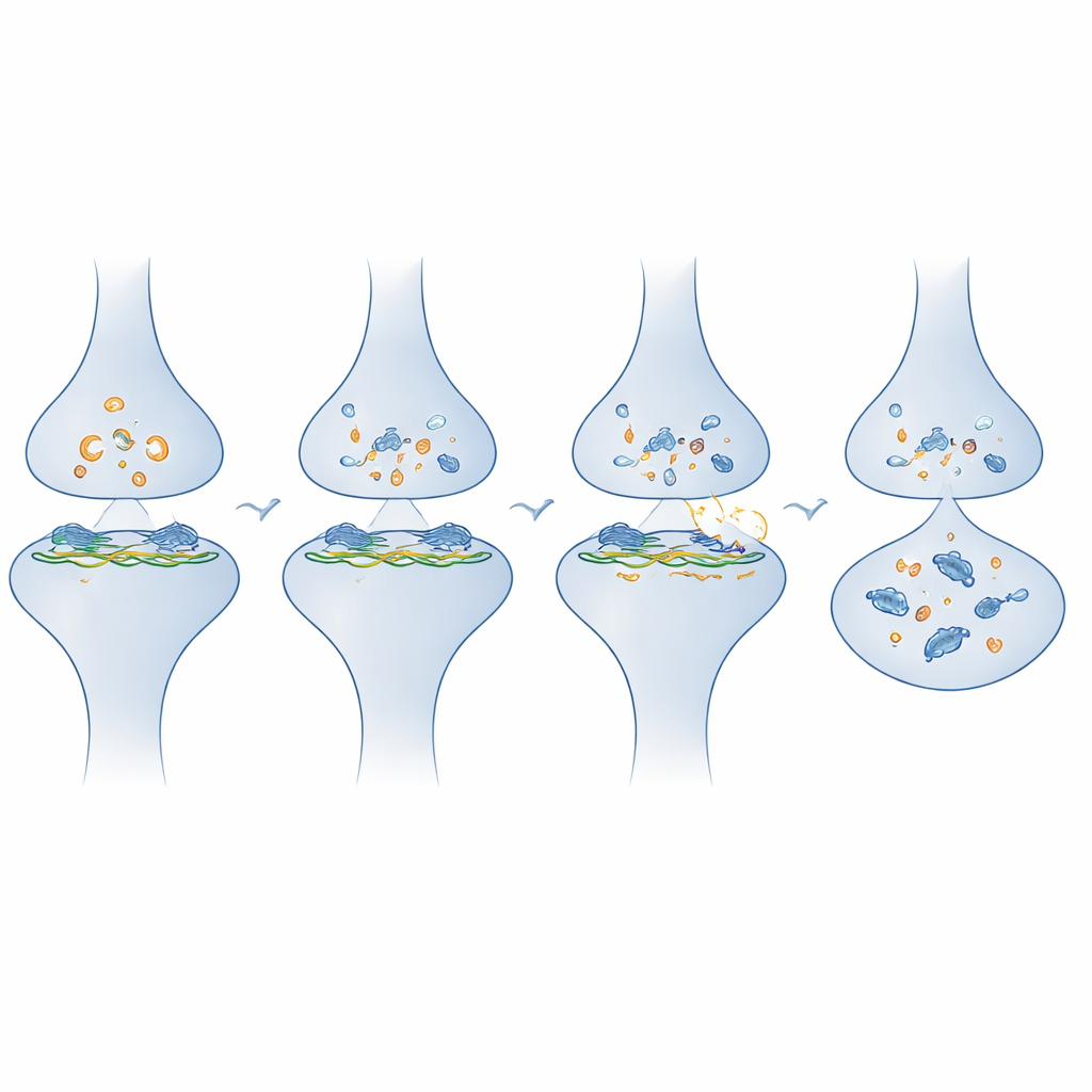
The standout finding was that CAP2 levels were clearly higher in people with motor neuron disease than in healthy volunteers, while SNAP-25 levels were not different between the two groups. This suggests that the receiving end of the nerve connection, and its internal structural machinery, is particularly altered in this disease. CAP2 is involved in shaping the tiny spines on nerve cells where incoming signals arrive and in managing the actin “framework” that keeps these spines stable yet flexible. Its rise in the spinal fluid points to active remodeling or stress at these postsynaptic sites, even when other synaptic proteins do not show obvious changes.
How CAP2 Differs from Classic Damage Markers
The team also compared CAP2 with more established markers that signal nerve fiber breakdown (neurofilament light chain) and support-cell activation (GFAP), as well as tau proteins, which reflect changes in the internal skeleton of nerve cells. People with motor neuron disease showed higher levels of all these injury markers overall, but CAP2 behaved differently. It did not track with neurofilament or GFAP, meaning it was not just mirroring general nerve or support-cell damage. Instead, CAP2 rose together with tau proteins only in patients, hinting at a shared disturbance in the cell’s structural systems that is specific to disease. Importantly, even after accounting for neurofilament levels, CAP2 still helped distinguish patients from healthy people, suggesting that it provides unique information about what is happening at synapses.
What These Signals Say About Disease Course
When the researchers followed patients over one year, they found that high neurofilament levels at the start predicted faster worsening of symptoms and poorer survival, confirming neurofilament as a strong marker of how aggressive the disease is. CAP2, on the other hand, did not predict how quickly the disease progressed or how long patients lived. It was consistently elevated across different clinical forms of motor neuron disease and across levels of symptom severity. This pattern suggests that CAP2 is less about how fast the disease advances and more about the presence of ongoing synaptic and structural rearrangement that accompanies the illness.
What This Means for Patients and Future Treatment
In plain terms, this study suggests that motor neuron disease is not just a story of dying nerve cells; it is also a story of stressed and reshaping connections between those cells. CAP2 appears to act as a window into these hidden changes at the receiving side of synapses and in the cell’s inner scaffolding, separate from the usual signals of nerve fiber breakdown. While CAP2 alone will not tell doctors how quickly a person’s disease will worsen, adding it to panels of other markers could give a more complete picture of the biology at work and help define subtypes of the disease. In the long run, such markers may guide therapies aimed at stabilizing synapses and preserving communication in the nervous system for as long as possible.
Citation: Pilotto, A., Pelucchi, S., Trasciatti, C. et al. Synaptic and cytoskeletal CSF signatures of motor neuron disease: the role of cyclase-associated protein 2. Sci Rep 16, 8703 (2026). https://doi.org/10.1038/s41598-026-39274-0
Keywords: motor neuron disease, ALS, cerebrospinal fluid biomarkers, synaptic dysfunction, CAP2 protein