Clear Sky Science · en
MultiScale hierarchical attention network for stain free breast cancer detection in microscopic hyperspectral imaging
Why looking at colorless tissue could change cancer care
Most hospital labs still rely on dyes and the expert eye of a pathologist to diagnose breast cancer. This study explores a different path: reading tiny fingerprints of light from completely unstained tissue and letting an artificial intelligence system decide whether cancer is present. If such a stain-free, automated approach proves reliable, it could shorten waiting times for results, reduce costs, and make diagnoses more consistent from one hospital to another.
Seeing more than the eye can see
Instead of the familiar pink and purple microscope slides, the researchers use microscopic hyperspectral imaging, which records how each point in a tissue slice reflects hundreds of precise colors of light. These spectra carry clues about the molecules inside the cells, such as proteins and nucleic acids, even though the tissue appears almost colorless to the naked eye. The team built a new dataset from 60 breast cancer patients, capturing 468 tissue sections. Each section was sampled at 20 locations, yielding three-dimensional data blocks that encode both fine cellular structure and rich color signatures far beyond standard red–green–blue images. 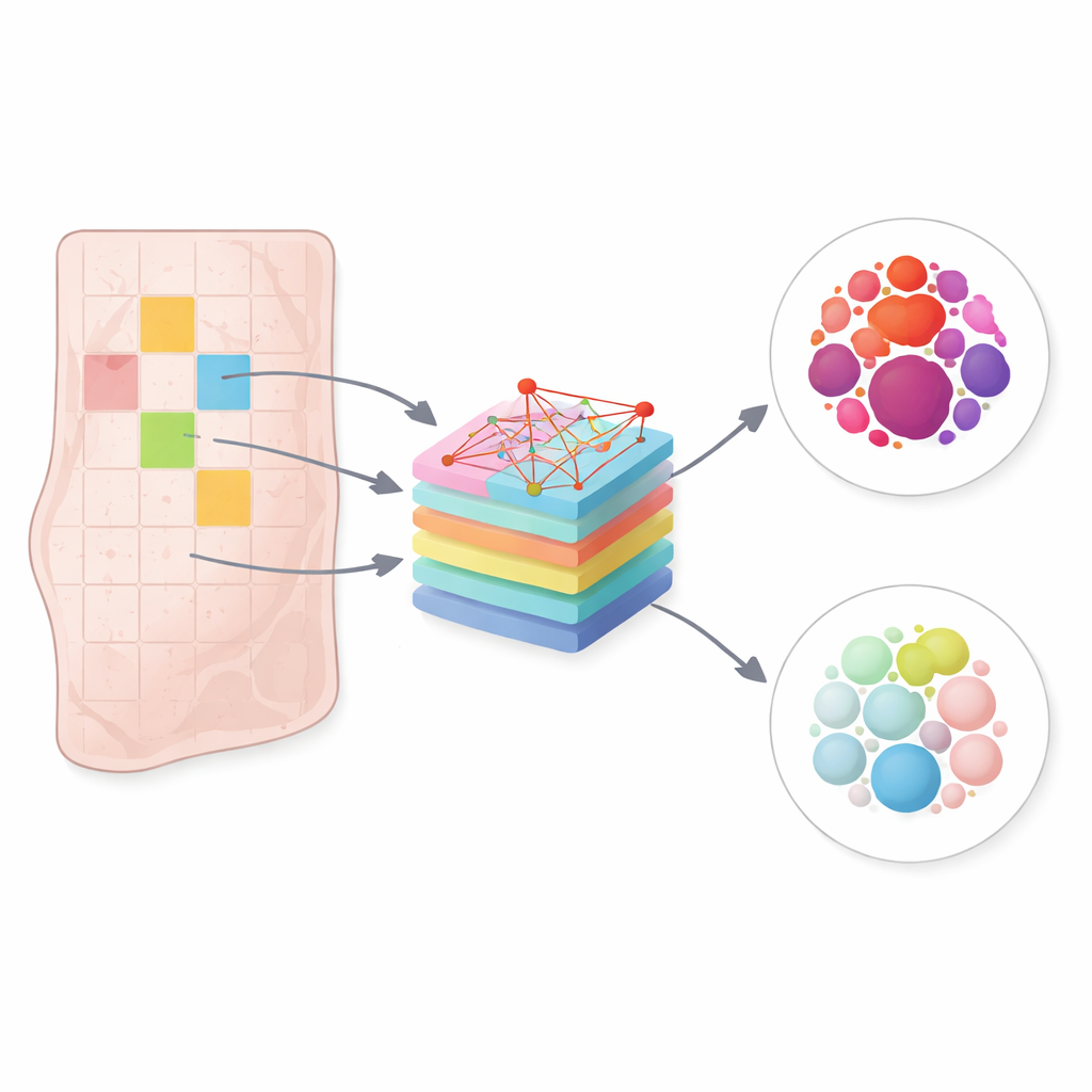
Letting the computer judge the whole slide
A major obstacle is that these stain-free images have weak visual contrast, and the distinctive cancer patterns are easily drowned out by noise and normal tissue. Rather than judging tiny regions in isolation, the authors reframed diagnosis as a “bag-level” decision: each tissue slice is treated as a collection of patches, and the model must weigh all of them together to decide whether the slice is cancerous or not. This setup, known in machine learning as multiple instance learning, mirrors how a pathologist mentally integrates clues from many fields of view before writing a report.
A smart spotlight on cells and colors
The core of the study is a new model called the MultiScale Hierarchical Attention Network (MS-HAN). For each patch, MS-HAN first uses several parallel filters of different sizes to capture patterns at multiple detail levels, from tiny cell features to slightly larger structures. It then applies a dual “attention” mechanism: one part learns which wavelengths of light are most informative, and another part highlights the most suspicious regions within the patch, much like a spotlight sweeping across the field of view. A built-in clustering step encourages patches with similar spectral fingerprints to group together around learned prototypes, making the model less sensitive to natural variation among different patients.
Piecing patches together into a final verdict
Once each patch has been distilled into a compact description, MS-HAN uses a transformer-like module to capture how patches relate to one another across the tissue slice. Some patches may reinforce each other’s signals, while others provide important contrast by looking more normal. An attention-based pooling step then combines these patch-level signals into a single portrait of the entire slice, which feeds into two coordinated decision branches that jointly produce the final cancer or non-cancer label. This layered, context-aware design aims to mimic how experts move from individual cell clusters to an overall judgment. 
How well does it work in practice?
On an unseen test set of 94 tissue slices, MS-HAN correctly distinguished tumor from nearby non-tumor tissue in about 87 out of 100 cases, with strong ability to avoid both missed cancers and unnecessary alarms. It outperformed several leading alternative methods that had been successful on conventional color slides, suggesting that tuning the architecture to the special demands of hyperspectral data pays off. Attention maps showed that the model focused on dense clusters of abnormal cells and on particular ranges of wavelengths, aligning qualitatively with what pathologists expect, although formal expert review of these visual explanations is still needed.
What this could mean for future patients
The study’s message is that stain-free breast cancer diagnosis using rich light spectra and a tailored attention-based model is technically feasible and can reach accuracy levels comparable to today’s best computer tools for stained slides. If validated in larger, multi-hospital cohorts and streamlined for speed, this approach could cut out chemical staining steps, speed up decisions during surgery, and offer more objective second opinions. In the long run, it hints at a future where a simple, label-free scan of tissue, interpreted by specialized AI, supports pathologists in delivering faster and more consistent cancer diagnoses.
Citation: Chen, Z., Yang, Q., Qin, G. et al. MultiScale hierarchical attention network for stain free breast cancer detection in microscopic hyperspectral imaging. Sci Rep 16, 9404 (2026). https://doi.org/10.1038/s41598-026-39267-z
Keywords: breast cancer diagnosis, hyperspectral imaging, stain-free pathology, deep learning attention, multiple instance learning