Clear Sky Science · en
Motion-robust myelin imaging in MRI using 1D projection gating
Why clearer brain scans matter
Doctors and scientists increasingly rely on MRI scans to see the brain’s wiring, especially the fatty coating called myelin that helps nerve signals travel quickly and reliably. Subtle changes in myelin are linked to conditions such as multiple sclerosis, concussion, epilepsy, and Alzheimer’s disease. But the MRI method that can see myelin most directly is slow and extremely sensitive to head motion, which makes it hard to use in everyday clinical practice—especially for patients who cannot stay perfectly still. This study introduces a way to make these delicate myelin scans much more tolerant of motion, without adding extra scan time or new hardware.
A hidden layer that powers brain speed
Myelin is a thin insulating sheath that wraps around nerve fibers in the brain and spinal cord. By allowing electrical signals to “jump” between gaps in the coating rather than crawl along the entire fiber, myelin boosts signaling speed by roughly a hundred-fold and greatly increases the brain’s information-carrying capacity. When myelin is damaged or lost, nerve signals slow down or fail altogether, contributing to problems with movement, vision, memory, and thinking. Standard MRI scanners, however, mostly see water in and around cells. Because the signal from myelin itself fades in a tiny fraction of a millisecond, and because the surrounding water is 10–20 times brighter, myelin is effectively invisible in routine scans.
A special MRI tuned to myelin
To tackle this, researchers have developed an advanced method called inversion-recovery ultrashort-echo-time (IR-UTE) imaging. It uses a carefully timed magnetic pulse to temporarily suppress the bright water signal, then listens almost immediately for the faint, rapidly disappearing signal from myelin. Two echoes are captured in quick succession and subtracted, so that lingering water contributions cancel out and the remaining image is strongly weighted toward myelin. This approach has already shown promise in tracking myelin loss in head injury and multiple sclerosis. The catch is that IR-UTE scans are long—around 10 minutes—and the resulting images are fragile: even small head movements can produce streaks and blurring that overwhelm the weak myelin signal.
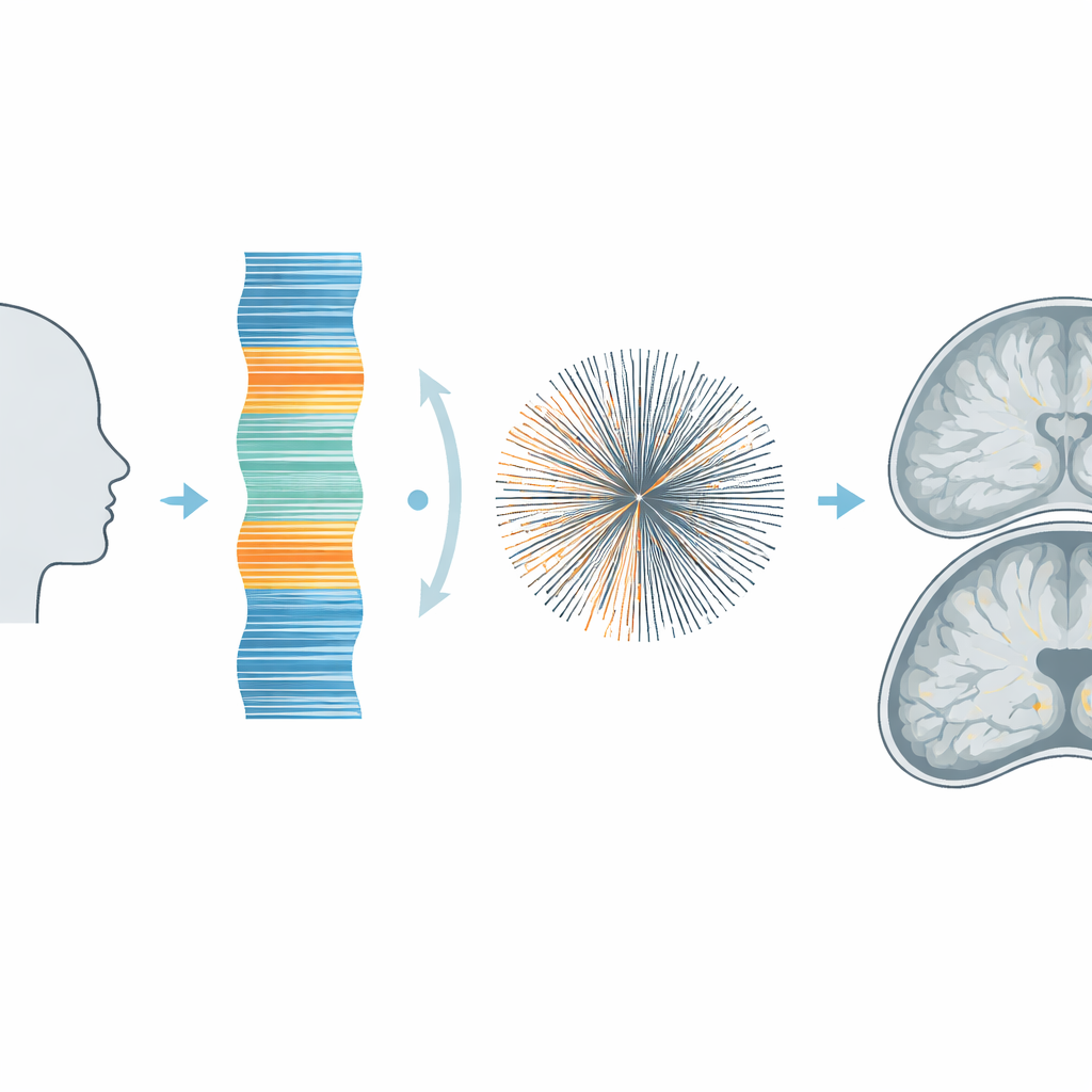
Listening for motion inside the scan
Instead of asking patients to hold still perfectly or adding cameras and extra sensors, the team designed a way for the MRI scan to monitor motion using its own data. At the end of each short imaging block, the scanner quickly measures how much signal comes from each level of the head along a single vertical line from top to bottom. This one-dimensional “shadow” of the head changes whenever the person nods or shifts. By comparing these profiles over time, the system identifies which segments of data were acquired during motion. Those corrupted pieces can then be excluded from the final image, a strategy known as retrospective gating—all done without lengthening the time between the main imaging pulses.
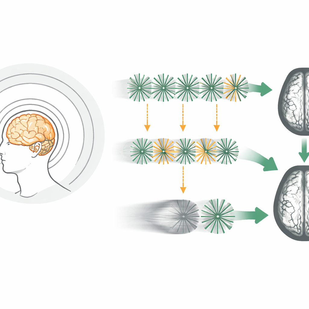
Scrambling the pattern to tame artifacts
Simply throwing away data that were acquired during motion can itself create new problems if all of the discarded measurements cluster in one region of the scanner’s sampling pattern. To avoid this, the researchers changed the order in which the scanner collects its radial “spokes” of data, using a mathematical trick called bit-reversed ordering. This reorders the spokes into a pseudo-random pattern so that, when 10 percent or more are rejected, the gaps are scattered evenly rather than forming a big missing wedge. Computer simulations with a digital brain model showed that the usual sequential ordering led to obvious streaks and blurred myelin-rich areas after gating, while the bit-reversed ordering produced much cleaner images with only low-level background noise.
Sharper myelin maps in real people
The team then tested their strategy on three healthy volunteers in a clinical 3‑tesla MRI scanner. They compared standard and bit-reversed spoke orderings, both without motion and with deliberate head nods during the scan. A simple threshold applied to the vertical motion signal identified about 11 percent of the data as motion-contaminated. When these data were removed, images acquired with conventional ordering lost contrast and showed patchy myelin signal, whereas the bit-reversed scans preserved fine detail in deep white matter and cortex. In scans with intentional motion, the gated, bit-reversed images were actually sharper and had better myelin-to-background contrast than images reconstructed from the full, ungated dataset, because blurring and ghosting from motion were largely suppressed.
Bringing motion-tolerant myelin MRI closer to the clinic
The study shows that combining an internal motion monitor with a smarter sampling pattern can turn a motion-sensitive, research-grade myelin scan into a more robust tool suitable for everyday use. By using a quick one-dimensional projection to detect when the head moves, and a bit-reversed ordering to spread any missing data evenly, the method improves myelin image quality without extra scan time or specialized hardware. In the future, this could make it easier to map myelin reliably in children, older adults, and patients with neurological disorders—opening a clearer window onto the brain’s wiring in situations where holding perfectly still is simply not possible.
Citation: Park, J., Sedaghat, S., Oguz, K.K. et al. Motion-robust myelin imaging in MRI using 1D projection gating. Sci Rep 16, 7866 (2026). https://doi.org/10.1038/s41598-026-39238-4
Keywords: myelin imaging, MRI motion correction, ultrashort echo time, brain white matter, neurodegenerative disease