Clear Sky Science · en
Three-dimensional reconstruction of a biliary system in a bioengineered liver using decellularized scaffold
Why Building New Livers Matters
Liver failure kills millions of people each year, and for many of them the only hope is a transplant. But donor livers are scarce, leaving patients waiting and often dying before an organ becomes available. Scientists therefore dream of growing replacement livers in the lab. A key missing piece has been a reliable way to rebuild the tiny tubes that carry bile—a fluid essential for digestion and waste removal—out of the liver. This study shows that it is now possible to rebuild much of that drainage system in a lab-made rat liver, bringing engineered replacement organs a step closer to reality. 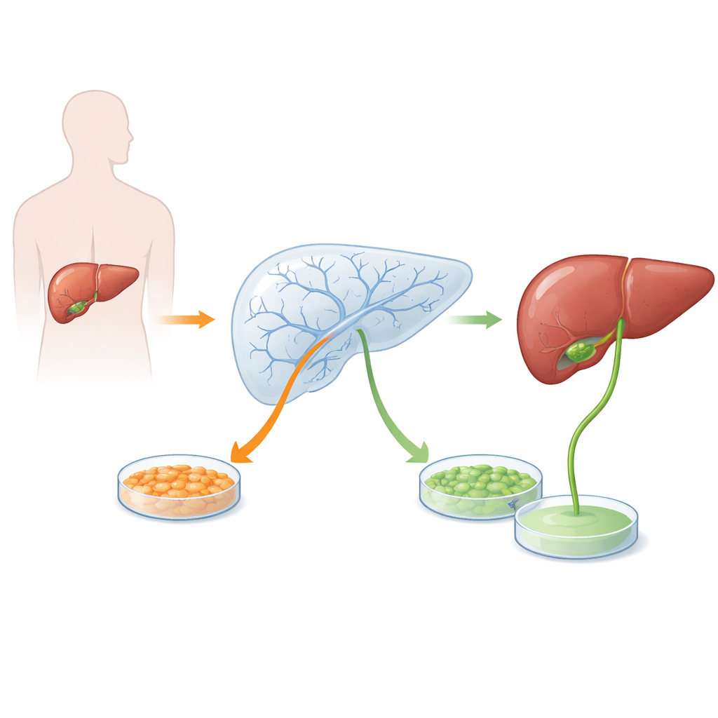
Turning a Real Organ into a Living Framework
The researchers began with whole rat livers and carefully removed all of the living cells, leaving behind only the nonliving support material known as the scaffold. This process, called decellularization, stripped away nuclei and other cell parts but preserved the fine tree of blood vessels and bile ducts that thread through the organ. The resulting translucent structure functioned like a detailed 3D mold of a liver, complete with hollow channels that could later be repopulated with new cells.
Seeding the Scaffold with Liver and Duct Cells
Next, the team introduced two types of rat cells into this empty framework. First were primary hepatocytes—the main workhorse cells of the liver that make bile, detoxify drugs, and produce many blood proteins. Second were intrahepatic cholangiocyte organoids, miniature clusters grown from bile duct fragments that behave like the cells lining bile ducts. The organoid cells were infused through the bile ducts and allowed to settle and grow for several days in a nutrient-rich, continuously perfused culture system. After that, hepatocytes were added, and the whole construct was cultured further so that both cell types could organize themselves within the scaffold. 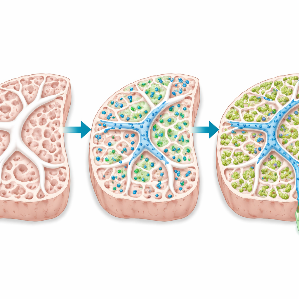
Rebuilding Tiny Bile Channels in Three Dimensions
Detailed microscopy showed that the organoid-derived cells successfully lined the inner surfaces of the preserved bile ducts, forming continuous tubular structures reminiscent of natural ducts. The hepatocytes spread through the surrounding tissue spaces and attached to the scaffold and to one another. Importantly, they re-established their internal polarity and formed narrow channels between neighboring cells known as bile canaliculi, through which bile normally first appears. In some regions, these newly formed canaliculi sat directly next to the rebuilt ducts, closely mimicking the arrangement seen in a healthy liver where bile flows from canaliculi into ducts and then out of the organ.
Hints That the New System Can Move Bile
To test whether this reconstructed network did more than just look right, the team measured bile acids—key components of bile—in liquid collected from the bile duct outlet and from the culture fluid circulating through the organ. In samples where the microscopic canaliculi and ducts were found close together, bile acid levels tended to be higher in the bile duct drainage than in the surrounding culture medium. This pattern is what one would expect if bile were being produced by hepatocytes, funneled into canaliculi, and then concentrated into the ducts. Although the sample size was small and the measurements preliminary, they offer early functional support for the idea that the rebuilt pathways can actually move bile.
Steps Toward Lab-Grown Replacement Livers
For non-specialists, the main takeaway is that scientists have managed to recreate much of the liver’s intricate bile drainage system inside an engineered organ, using a real liver framework repopulated with two carefully chosen cell types. The work does not yet produce a fully functional transplant-ready liver, and many challenges remain, including improving cell survival, achieving more uniform rebuilding throughout the organ, and proving robust bile flow over time and in living animals. Still, this research demonstrates that the 3D architecture needed for bile transport can be reassembled, moving the field of bioengineered livers closer to organs that could one day replace damaged livers in patients.
Citation: Horie, H., Fukumitsu, K., Hanabata, Y. et al. Three-dimensional reconstruction of a biliary system in a bioengineered liver using decellularized scaffold. Sci Rep 16, 8071 (2026). https://doi.org/10.1038/s41598-026-39175-2
Keywords: bioengineered liver, bile ducts, tissue engineering, organ scaffolds, liver regeneration