Clear Sky Science · en
Machine learning-enhanced multi-band metamaterial sensor for early detection of neurological disorders
Seeing Brain Trouble Before Symptoms Appear
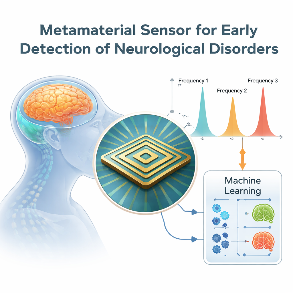
Neurological disorders such as brain tumors, multiple sclerosis, and traumatic injuries often begin with subtle changes that today’s scanners can miss. This study introduces a tiny sensor, built from specially patterned materials and aided by machine learning, that operates with terahertz light to spot early shifts in the brain’s fluids and tissues. In the future, such a chip could help doctors detect problems sooner, using smaller and potentially cheaper equipment than today’s bulky MRI or CT machines.
Why a New Kind of Brain Sensor Is Needed
Doctors currently rely on CT and MRI to locate damaged brain tissue, but these machines are large, expensive, and not always sensitive to the earliest stages of disease. Many brain conditions subtly alter the properties of cerebrospinal fluid—the clear liquid that cushions the brain and spinal cord and helps keep them chemically stable. When its water content changes, so does the way it bends light, a quantity known as the refractive index. Conventional scanners are not designed to measure these tiny optical changes directly. The authors argue that if a compact sensor could read these shifts with high precision, it might reveal signs of trouble long before structural damage becomes obvious.
Harnessing Terahertz Light and Designer Materials
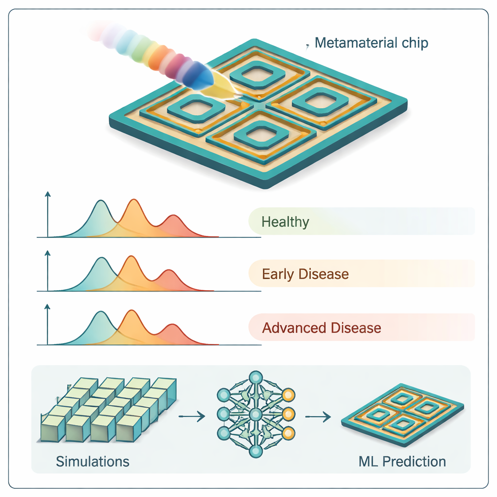
The proposed sensor works in the terahertz region of the electromagnetic spectrum, a band of radiation that can penetrate biological tissue without the harmful ionizing effects of X-rays. At the heart of the device is a carefully patterned “metamaterial” surface: a 35-micrometer-wide square made from gold and a plastic called polyimide, arranged as nested square and octagonal loops. Rather than relying on the raw composition of the materials, the design uses geometry to trap incoming terahertz waves very efficiently. When the sensor is exposed to a sample—such as cerebrospinal fluid or brain-like tissue—its absorption spectrum shows three very sharp peaks at specific frequencies. Because more than 99 percent of the incoming terahertz energy is absorbed at each peak, small shifts in those frequencies become easy to detect.
Reading Tiny Changes in Brain-Like Tissues
To test its sensing power, the team placed a thin “analyte” layer above the metamaterial and varied its refractive index across the range typical of biological fluids. Each time the refractive index changed, all three absorption peaks moved to slightly different frequencies while staying very strong, above about 96 percent absorption. From these shifts, the researchers calculated sensitivity values of 1.5, 1.5, and 1.8 terahertz per refractive-index unit for the three peaks—figures that compare favorably with or exceed many earlier terahertz sensors. They then modeled realistic brain conditions by assigning refractive index values to different tissues, including healthy cerebrospinal fluid, gray and white matter, and several kinds of brain tumors. The three resonance peaks for each tissue type separated cleanly without overlapping, indicating that the device could, in principle, distinguish between healthy and diseased states across multiple channels at once.
Speeding Up Design with Machine Learning
Designing such a finely tuned sensor usually demands thousands of time-consuming computer simulations. To overcome this, the authors generated a large dataset by systematically varying five key design parameters—such as layer thicknesses and gap sizes—and recording the resulting absorption. They then trained several machine learning models to predict the sensor’s response without running full simulations. Gradient boosting, a popular ensemble method, emerged as the top performer, reproducing the simulated absorption curves with extremely high accuracy. By relying on these learned models, the team estimates they can explore new designs while cutting simulation time by up to 60 percent. They further used explainable AI tools, SHAP and LIME, to identify which parameters mattered most, offering insight into how geometry controls sensing performance.
What This Could Mean for Early Diagnosis
In simple terms, the study shows that a postage-stamp-sized terahertz chip can act as a very sharp “ear” for listening to how brain fluids and tissues interact with light, and that these interactions change in reliable ways as disease progresses. Because the sensor produces three independent readings at once, it gains both accuracy and robustness: if one channel is disturbed, the others can still help identify the tissue state. While the work so far is based on simulations and needs to be confirmed in laboratory and clinical settings, the combination of high sensitivity, compact size, and machine-learning-guided design suggests a promising route toward faster, more accessible tools for catching neurological disorders at their earliest, most treatable stages.
Citation: Miah, A., Al Zafir, S., Das, J. et al. Machine learning-enhanced multi-band metamaterial sensor for early detection of neurological disorders. Sci Rep 16, 7599 (2026). https://doi.org/10.1038/s41598-026-39127-w
Keywords: neurological disorders, terahertz sensing, metamaterial sensor, cerebrospinal fluid, machine learning