Clear Sky Science · en
SwinCup-DiscNet: A fusion transformer framework for glaucoma diagnosis using optic disc and cup features
Why this matters for saving sight
Glaucoma is one of the world’s leading causes of irreversible blindness, yet it often creeps up silently, without pain or early warning signs. Eye doctors can spot subtle changes in the back of the eye before vision is lost, but doing this by hand for every patient is slow and sometimes inconsistent. This paper introduces SwinCup-DiscNet, a new artificial intelligence (AI) system that reads retinal photographs to flag glaucoma early, combining classic clinical cues with modern deep learning. 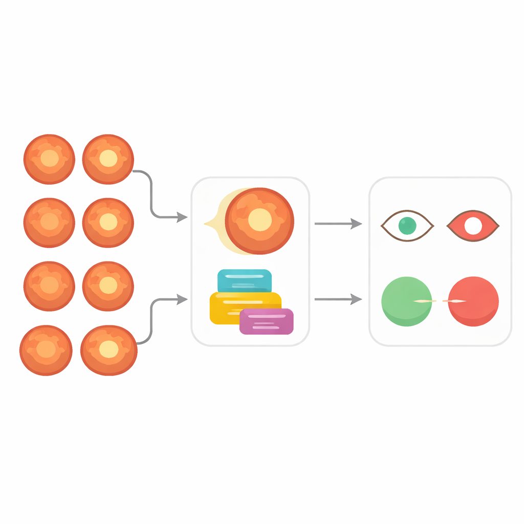
Looking at the nerve inside the eye
To understand what the system does, it helps to know how glaucoma is usually spotted. Eye specialists examine the optic nerve head, the spot where the nerve that carries visual information leaves the eye. In the center of this “disc” is a lighter depression called the “cup.” As glaucoma progresses, the cup tends to deepen and widen, eating into the surrounding rim of nerve tissue. A key number is the cup-to-disc ratio, which compares the size of the cup to the size of the disc. A higher ratio often signals damage. Measuring this ratio by hand on thousands of retinal photographs is tedious, and even experts can disagree. SwinCup-DiscNet automates both the measurement of this ratio and the overall judgment about whether an eye is likely to have glaucoma.
A two-track AI that sees details and the bigger picture
The system follows two parallel tracks when it receives a retinal fundus image. First, a segmentation branch isolates the optic disc and the central cup. It uses a specialized network known as Attention U-Net, which learns to highlight the important structures and ignore distracting background features like blood vessels and lighting artifacts. Once it has identified the cup and disc boundaries, the system smooths them and fits clean oval shapes, then measures their vertical sizes to compute the vertical cup-to-disc ratio—a clinically trusted marker of glaucoma.
Learning patterns beyond what the eye can measure
In the second track, a transformer-based branch looks at the entire image without focusing on any single number. This branch uses a Swin Transformer, a modern deep learning model that divides the image into small patches and analyzes how they relate to each other across the whole retina. In doing so, it picks up subtle patterns in texture, color, and structure around the optic nerve and nearby regions that might be linked to glaucoma but are hard for humans to quantify. From this global view, the model produces a probability that the image comes from a person with glaucoma. 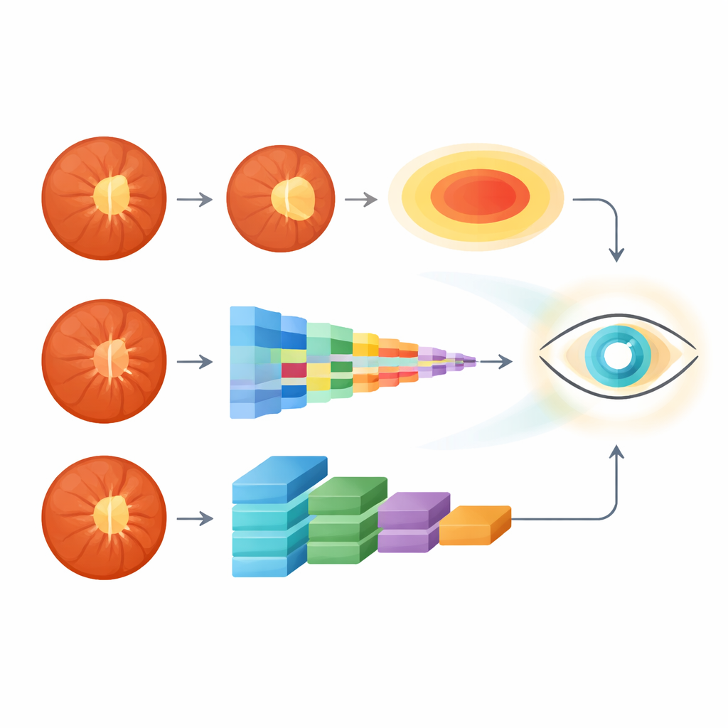
Blending trusted clues with AI intuition
The heart of SwinCup-DiscNet is how it merges these two sources of evidence. Instead of trusting only the cup-to-disc ratio or only the transformer’s probability, the system blends them using a weighted rule. The cup-to-disc ratio is normalized based on how it behaved in training data, then combined with the model’s learned glaucoma probability into a single score. If that fused score passes a threshold, the eye is classified as glaucomatous; if not, it is labeled normal. This design keeps the decision anchored in a familiar clinical measurement while still taking advantage of the richer patterns the AI can detect. The system also overlays the fitted disc and cup outlines on the original image, giving doctors a clear visual of which region drove the decision.
Putting the method to the test
The authors evaluated SwinCup-DiscNet on three widely used public datasets of retinal images: LAG, ACRIMA, and DRISHTI-GS. These collections vary in camera type, image quality, and patient mix, making them a tough test bed. Across all of them, the new system matched or outperformed traditional convolutional networks and methods that only segment the cup and disc. It achieved very high segmentation quality, low error in estimating the cup-to-disc ratio, and classification accuracies close to or above 99 percent, with strong performance curves indicating it rarely confuses healthy and diseased eyes. An analysis of errors showed that most remaining false alarms were in borderline cases where the optic cup was naturally large but not truly diseased, a trade-off that is often acceptable in screening.
What this means for future eye screening
In plain terms, SwinCup-DiscNet shows that AI can both “think like a doctor” by using established markers such as the cup-to-disc ratio and “see beyond the obvious” by learning complex patterns in retinal images. By combining these strengths, the system delivers accurate and more interpretable glaucoma screening than many existing approaches. With further testing on real-world hospital data and possible extensions to grade disease severity, this kind of hybrid AI could become a practical assistant in eye clinics, helping catch glaucoma earlier and prevent avoidable blindness.
Citation: Chilukuri, R., Praveen, P., Gatla, R.K. et al. SwinCup-DiscNet: A fusion transformer framework for glaucoma diagnosis using optic disc and cup features. Sci Rep 16, 7920 (2026). https://doi.org/10.1038/s41598-026-39065-7
Keywords: glaucoma, retinal imaging, deep learning, optic nerve, medical screening