Clear Sky Science · en
A numerical flow experiment for assessing the risk of rupture in anterior communicating artery aneurysms in relation to aneurysm projection
Why the Shape of a Tiny Bulge in the Brain Matters
Brain aneurysms are small bulges in blood vessels that can sit silently for years yet, if they burst, cause life‑threatening bleeding. Doctors often judge how dangerous an aneurysm is by its size, but many ruptures still occur in bulges considered “too small to worry about.” This study asks a deceptively simple question with serious consequences: does the direction in which an aneurysm points change how blood flows inside it, and therefore how likely it is to burst or to slowly fill with clot?
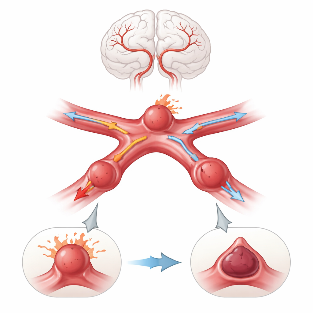
A Closer Look at a Problematic Junction
The work focuses on aneurysms in one of the brain’s riskiest spots: the anterior communicating artery, a tiny connection between major vessels at the base of the brain. Aneurysms in this location can balloon toward the front of the head, back toward the optic nerves, upward toward deep brain structures, or downward toward the skull base. Rather than studying only real‑world scans, the researchers built a detailed but idealized 3D model of the main brain arteries, including a complete circle of Willis. Onto this virtual vessel network, they attached aneurysm domes that were identical in size and neck shape but pointed in four different directions. They then created both “small” and “large” versions of these bulges to see whether size changed the flow story.
Simulating Blood Flow Beat by Beat
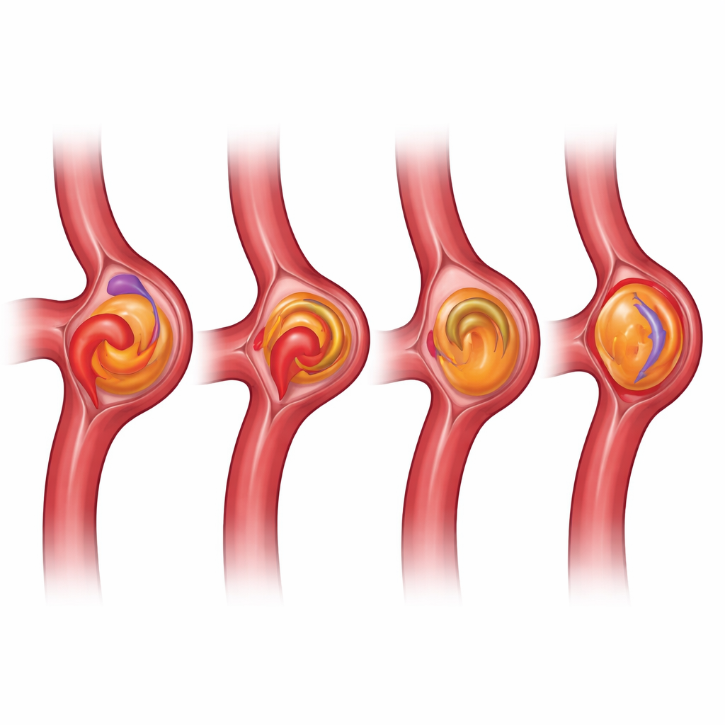
To explore how blood behaves inside each aneurysm type, the team turned to computer fluid simulations similar to those used in aerodynamics. They modeled blood as a realistic, thick‑but‑flowing liquid whose consistency changes with speed, and drove it through the arteries using a pulsing pattern that mimics a human heartbeat. The virtual heart pumped for ten full cardiac cycles. At the inlets, they fed in “new” blood while tracking how quickly it pushed “old” blood out of the aneurysm sac. This allowed them to measure not just pressure and wall stress, but also how long blood tended to linger in each bulge, how strongly it swirled, and how effectively it was rinsed clean with every beat.
Fast Rivers, Slow Ponds, and Hidden Dangers
The simulations revealed that pointing direction, not size, was the main architect of the internal flow patterns. When the aneurysm projected forward (anteriorly), blood shot into it with higher speed, formed strong swirling currents, and quickly washed back out, leaving almost no old blood after several heartbeats. The walls of these forward‑facing bulges experienced higher pressure and stronger rubbing forces, conditions that earlier work has linked to weakening of vessel tissue and a greater chance of rupture. In contrast, downward‑pointing (inferior) aneurysms behaved like slow ponds. Blood crept in sluggishly, swirled weakly, and large pockets of old blood remained even after many cycles. Inside these domes the fluid became thicker and more stagnant—an environment known to favor clot formation rather than sudden bursting. Upward and backward projections landed in between, with moderate flow speeds, stresses, and washout.
Same Story for Small and Large Bulges
One might expect a larger aneurysm to be much more unstable simply because of its size. Surprisingly, the overall patterns of flow and stress within each projection type hardly changed when the authors scaled the bulge up. Large anterior aneurysms still showed fast, energetic circulation and efficient washout, and large inferior aneurysms still trapped old, sluggish blood. Absolute values of pressure and flow increased, but the relative ranking of “lively and stressed” versus “quiet and stagnant” projections remained the same. This reinforces growing clinical evidence that many small aneurysms can be dangerous, and that shape and orientation may tell a richer story than diameter alone.
What This Means for Patients and Doctors
Seen through the lens of these simulations, two aneurysms of identical size at the same arterial junction can live very different lives depending on which way they point. A forward‑facing bulge is bathed in fast, swirling blood that may erode its wall and raise rupture risk, while a downward‑facing one is more likely to collect clot and remain stable but may pose challenges during treatment. Because these trends held for both small and large domes, the study argues that doctors should look beyond simple size thresholds and include projection‑specific flow information when deciding how closely to watch an aneurysm or when to intervene. In everyday terms, it is not just how big the bulge is, but how the blood runs through it, that may determine whether it quietly scars over or suddenly bursts.
Citation: Wiśniewski, K., Tyfa, Z., Dębska, A. et al. A numerical flow experiment for assessing the risk of rupture in anterior communicating artery aneurysms in relation to aneurysm projection. Sci Rep 16, 8317 (2026). https://doi.org/10.1038/s41598-026-38826-8
Keywords: brain aneurysm, blood flow simulation, cerebral circulation, rupture risk, aneurysm projection