Clear Sky Science · en
Hybrid EfficientNet B4 and SVM framework for rapid and accurate bone cancer diagnosis from X-rays
Why faster bone cancer checks matter
Bone cancer is rare but devastating, and spotting it early on X‑rays can be surprisingly difficult, even for experienced doctors. Subtle tumors can resemble harmless changes, and radiologists must carefully inspect hundreds of images, often under time pressure. This paper introduces a new computer-based assistant called OsteoCancerNet that aims to help doctors read bone X‑rays faster and more accurately, catching dangerous tumors while keeping false alarms low.
The problem with looking by eye alone
Doctors currently rely on imaging tools such as X‑rays, CT, and MRI scans to find bone tumors and plan treatment. But these pictures are still interpreted by humans, which introduces delays and the risk of missed or mistaken findings, especially when lesions are small or look similar to normal bone. Earlier research has shown that artificial intelligence can help analyze medical images, yet many systems for bone cancer have used small image collections, taken a long time to run, or behaved like “black boxes” that are hard to test and trust. Some models recognize patterns well but are too large and slow for everyday hospital use, while others work only on narrow, highly curated datasets.
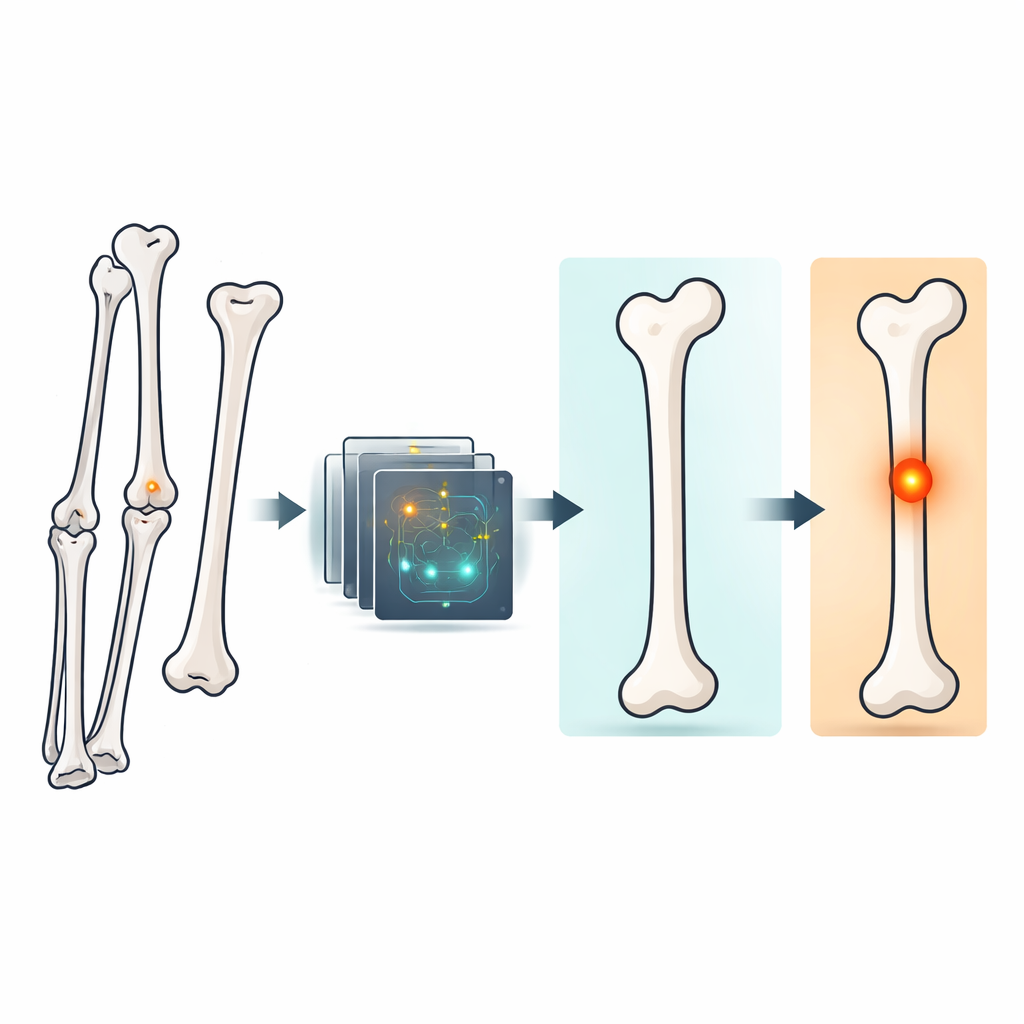
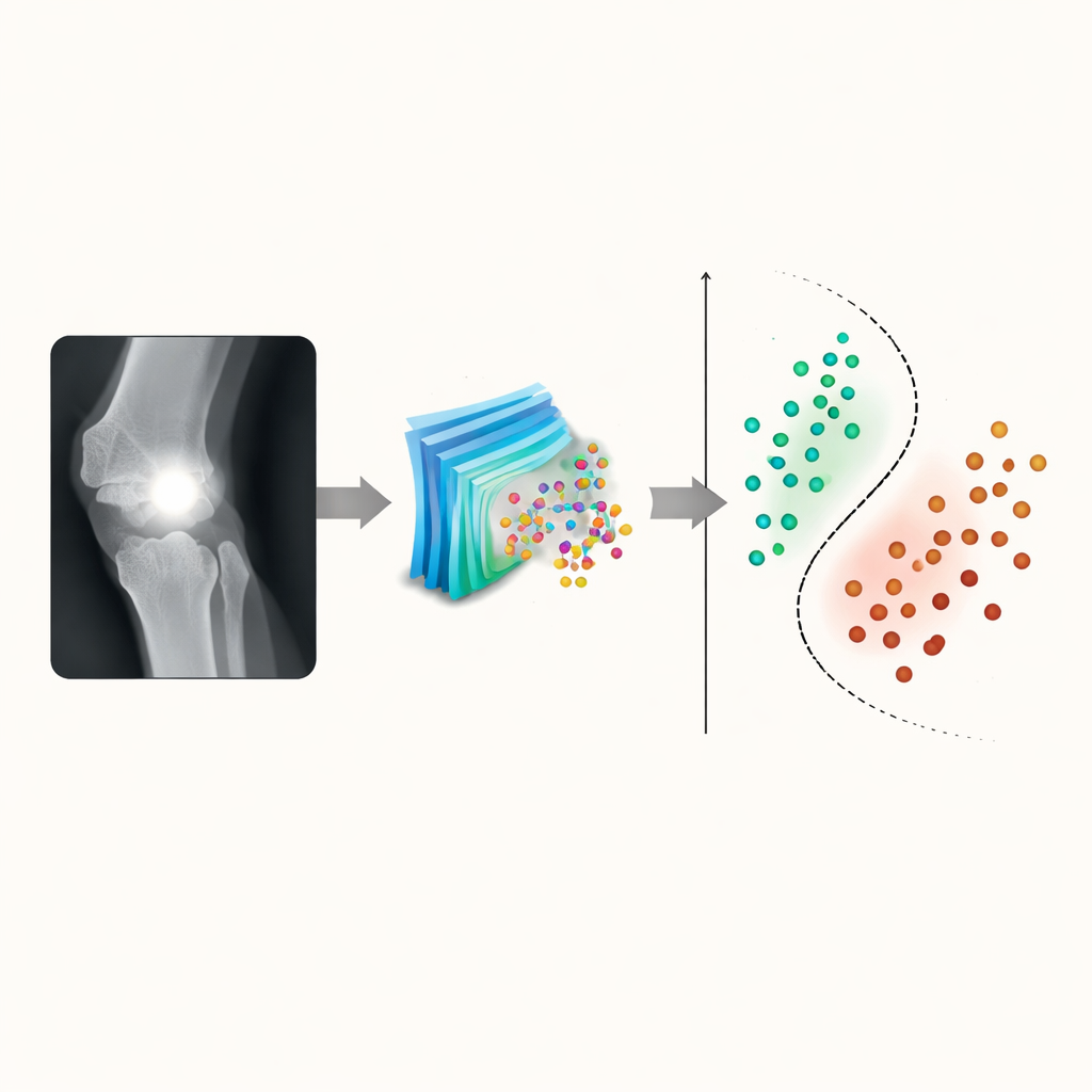
A smart blend of two AI approaches
OsteoCancerNet combines two complementary AI tools to get the best of both worlds. First, it uses a modern deep-learning network called EfficientNet-B4 to scan each bone X‑ray and automatically learn rich visual features—subtle changes in shape, texture, and contrast that may signal cancer. Instead of directly deciding from these raw features, the system then hands them to a more classical machine-learning method called a support vector machine, which acts as the final decision-maker, separating “normal” from “cancerous” images. This hybrid design is meant to capture complex image details while keeping the final classification step relatively simple, stable, and easier to evaluate.
Cleaning and multiplying the X‑ray data
To build and test the system, the researchers used a large public collection of 8,811 bone X‑ray images, evenly split between healthy and cancer cases. They first cleaned and standardized these images so the AI would see consistent inputs. Each X‑ray was resized to the required format, converted to color channels the network expects, and then sharpened through several contrast-enhancement methods. A technique called CLAHE, which selectively boosts contrast in local regions without washing out fine details, turned out to produce the clearest pictures for the AI. Because medical datasets are often small, the team also “augmented” the training images by flipping and rotating them, effectively growing the training set to nearly 30,000 images. This makes the system more robust to different viewing angles and reduces the risk of overfitting to a particular dataset.
How well the system detects bone cancer
After training, OsteoCancerNet was evaluated on several fronts. On a held‑out test set of X‑rays it had never seen, the model correctly classified about 97 out of 100 images and showed a strong balance between catching cancers and avoiding false alarms. Its overall accuracy was roughly 98% during cross‑validation, with a very high ability to detect true cancer cases and a very low false positive rate of about four in ten thousand normal images. Crucially, the system is fast: once trained, it needs only about 41 milliseconds to analyze a single X‑ray, fast enough for real‑time use in a busy clinic. The researchers also compared OsteoCancerNet with a wide range of other popular AI models, including well‑known deep networks and hybrid systems, and found that their approach consistently delivered higher accuracy with fewer mistaken alerts and more modest computing demands.
What this means for patients and doctors
The study shows that carefully crafted AI can act as a reliable second set of eyes for bone X‑ray reading. By sharpening images, using an efficient deep network to capture subtle bone changes, and delegating the final decision to a streamlined classifier, OsteoCancerNet spots bone cancer with impressive consistency and speed. For patients, this could translate into earlier detection, fewer missed tumors, and quicker reassurance when scans are normal. For clinicians, the system offers a practical tool that reduces workload rather than adding to it. While further testing in real-world hospital settings and across more imaging types is still needed, the work points toward AI‑assisted bone cancer diagnosis becoming a routine, trustworthy part of orthopedic and cancer care.
Citation: Hassan, N.M.H., Bayoumy, A.S. & Mahmoud, M.H.M. Hybrid EfficientNet B4 and SVM framework for rapid and accurate bone cancer diagnosis from X-rays. Sci Rep 16, 8156 (2026). https://doi.org/10.1038/s41598-026-38801-3
Keywords: bone cancer, medical imaging AI, X-ray analysis, deep learning, computer-aided diagnosis