Clear Sky Science · en
Intravenous high mobility group box 1 fragment improves cardiac function, fibrosis, and coronary flow in porcine ischemic cardiomyopathy model
Why this heart study matters
Heart failure is becoming more common as populations age, and many patients with severe damage from heart attacks eventually run out of treatment options. Operations like heart transplants or mechanical pumps can be lifesaving but are highly invasive and not suitable for everyone. This study explores a less invasive approach: a small fragment of a natural protein, given through a simple vein drip, that appears to help the heart repair itself in a large-animal model that closely resembles human heart disease.
A new way to tap the body’s own repair crew
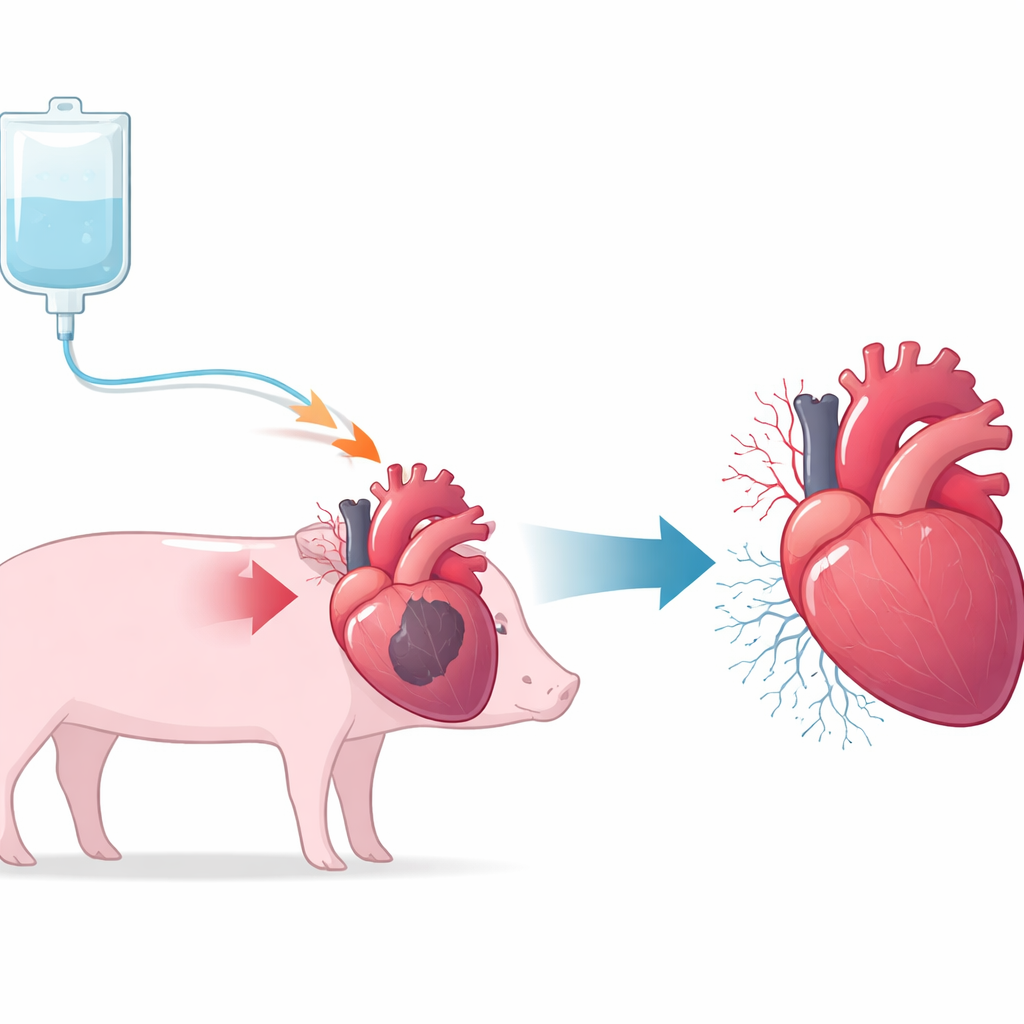
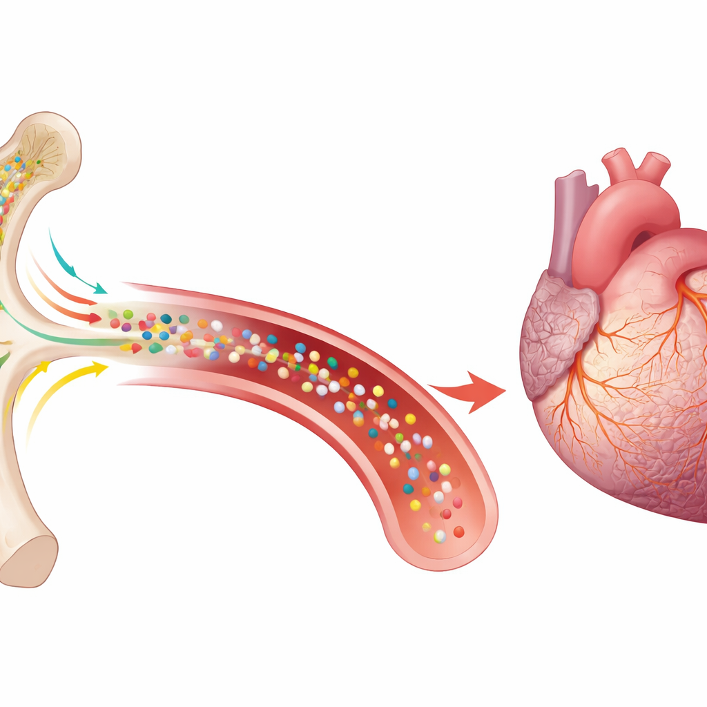
The researchers focused on a protein called high mobility group box 1, or HMGB1. In the body, HMGB1 plays roles in controlling genes, inflammation, and tissue healing. Earlier work showed that a specific fragment of HMGB1 can coax repair cells from the bone marrow—called mesenchymal stem cells—into the bloodstream and toward injured tissues. The team asked whether this fragment, delivered into a vein, could improve heart function in pigs with ischemic cardiomyopathy, a condition where past loss of blood flow leaves the heart weak and scarred, similar to many patients after heart attacks.
Building a realistic model of a damaged heart
To mimic chronic coronary artery disease, the scientists partially choked off a major heart artery in mini pigs using a ring that slowly tightens, creating long-lasting poor blood flow and scarring. After four weeks, the animals had weakened pumping function, enlarged hearts, and damaged regions visible on advanced scans. The pigs were then randomly split into two groups: one received five intravenous doses of the HMGB1 fragment over ten days, while the other received only saline. The animals were followed for another eight weeks with echocardiography, cardiac MRI, pressure measurements inside the coronary arteries, and detailed tissue analysis after humane euthanasia.
Stronger heartbeat, less scar, better blood flow
Across several types of imaging, the treated pigs showed a clear pattern of recovery compared with controls. Standard ultrasound and MRI both revealed that the heart’s pumping efficiency—the fraction of blood pushed out with each beat—increased in the HMGB1 group but not in untreated animals. The volume of blood left behind after each contraction shrank, signaling a stronger, more effective squeeze. MRI scans that highlight scar tissue showed that damaged, non-working muscle areas actually shrank in treated pigs, while they expanded in controls. Measurements taken with thin wires inside the coronary arteries indicated that the ability of the vessels to boost flow when needed—called coronary flow reserve—improved with HMGB1 treatment, suggesting healthier small vessels feeding the heart muscle.
Signs of healing under the microscope
When the hearts were examined directly, the border zones around the old injury looked healthier in the treated animals. Muscle cells were smaller and more uniform, rather than stretched and swollen as seen in failing hearts. There was a trend toward less fibrous scar tissue, and many more tiny blood vessels lined by specialized cells, consistent with new vessel growth. Molecular tests showed higher levels of several factors known to encourage vessel formation, limit scarring, and calm excessive inflammation. Markers linked to bone marrow–derived repair cells were also somewhat higher, supporting the idea that the drug fragment had recruited the body’s own cell-based repair system rather than acting as a conventional drug that targets a single pathway.
What this could mean for future patients
Taken together, the findings suggest that repeated intravenous doses of an HMGB1 fragment can trigger a broad self-repair program in a large-animal model of chronic heart damage. The treatment seemed to spur new blood vessel growth, reduce harmful scarring, and restore function in hibernating but still living heart muscle, all without the need to transplant cells or perform high-risk surgery. While more work is needed to confirm safety, refine dosing, and show benefit in people, this approach points toward a future in which some forms of severe heart failure might be treated by awakening the heart’s own capacity to heal.
Citation: Ito, Y., Kawamura, M., Kawamura, T. et al. Intravenous high mobility group box 1 fragment improves cardiac function, fibrosis, and coronary flow in porcine ischemic cardiomyopathy model. Sci Rep 16, 8350 (2026). https://doi.org/10.1038/s41598-026-38592-7
Keywords: heart failure, ischemic cardiomyopathy, regenerative therapy, mesenchymal stem cells, HMGB1 fragment