Clear Sky Science · en
Radiomic features and carotid stenosis in periodontitis a two stage bootstrap and multimodal machine learning study
Why your gums might say something about your heart
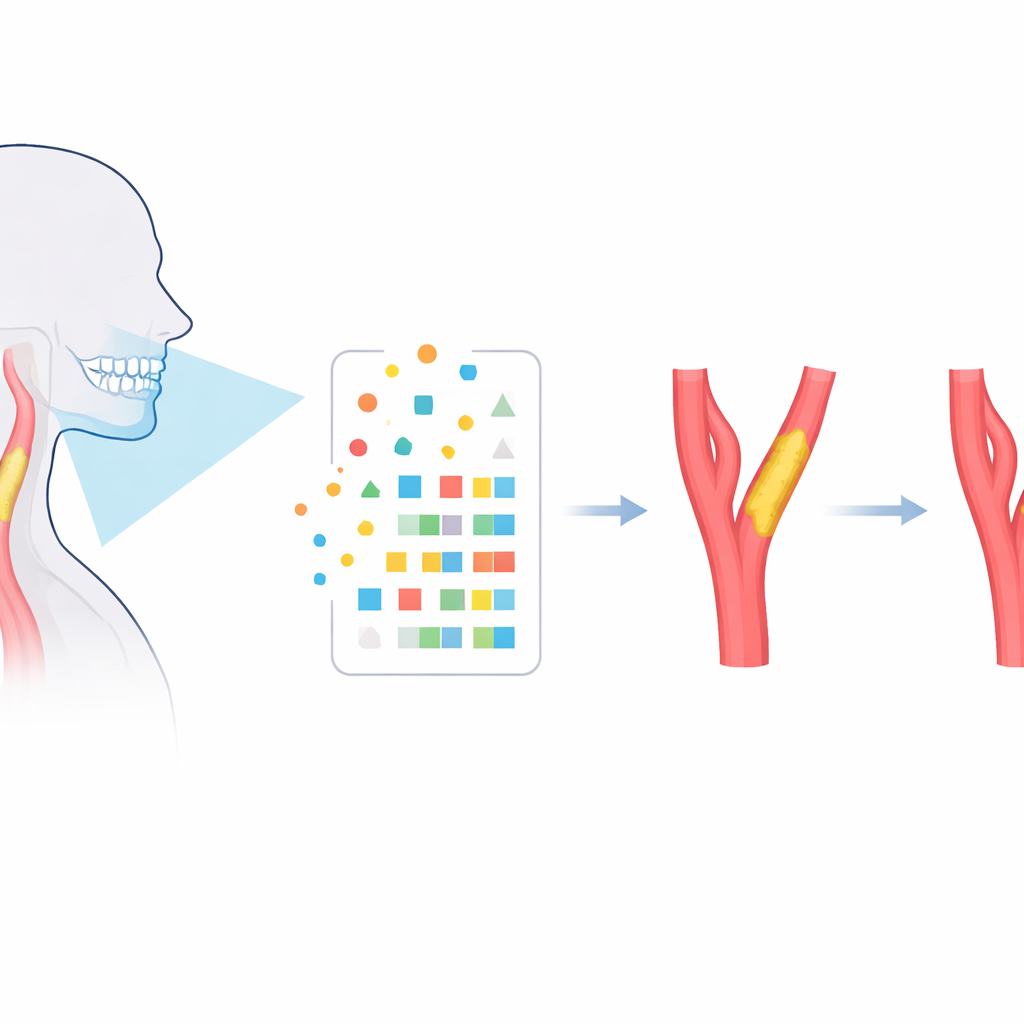
Most of us think of dental X-rays as tools for spotting cavities or planning implants. This study suggests they may also whisper clues about the health of the neck arteries that feed the brain. By mining hidden patterns in routine 3D dental scans from people with gum disease, the researchers show it may be possible to flag those at higher risk of dangerous artery narrowing long before a stroke occurs.
Hidden warning signs in the mouth
Gum disease, or periodontitis, is a long-lasting infection that slowly damages the tissues supporting our teeth. Over the past decade, many studies have linked it to heart attacks and strokes, hinting that inflamed gums and diseased blood vessels may be different faces of the same problem. Yet doctors still lack simple, practical tools to single out which patients with periodontitis are quietly developing carotid artery narrowing in the neck, a major cause of ischemic stroke. The authors asked whether the 3D cone beam CT scans already used in dental clinics might hold subtle structural clues that reflect this hidden artery damage.
Turning dental scans into measurable patterns
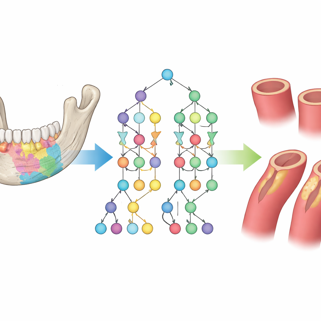
The team analyzed cone beam CT scans from 279 adults treated at a large hospital: 168 had both periodontitis and carotid artery narrowing, while 111 had periodontitis alone. For each person, specialists carefully outlined the upper and lower jaw regions that house the teeth and supporting bone. Using radiomics—a technique that converts medical images into lots of numerical descriptors—they extracted 206 features per scan. These included simple measures such as overall brightness, as well as shape and texture patterns that are impossible to judge by eye but may reflect how inflammation and bone remodeling have altered the jaw over time.
Teaching machines to spot at-risk patients
Because more patients in the study had diseased arteries than healthy ones, the researchers used a data-balancing method called SMOTE to create a more even training set for their algorithms. They then applied a careful, two-step statistical process to sift through the 206 radiomic features. First, they repeatedly resampled the data and used correlation checks and a shrinking regression method to discard redundant or weak signals. Features that repeatedly survived this gauntlet were carried into a second stage, where repeated logistic regression runs chose the most stable combination. This winnowing left 20 key features—covering jaw shape, intensity distribution, and fine-grained texture—that together best distinguished patients with and without carotid narrowing.
How well the models performed
With these 20 features, the team built and compared three common machine-learning models: logistic regression, support vector machines, and random forests. Using five-fold cross-validation—a way of testing performance on unseen data—they found that the random forest model worked best. It correctly separated high-risk from lower-risk patients with an area under the curve of 0.892, very high sensitivity (it caught about 96% of those with artery narrowing), and moderate specificity (it correctly reassured about 71% of those without it). Additional checks showed that its probability estimates matched reality reasonably well and that, across a wide range of decision thresholds, it would give clinicians more net benefit than either the simpler models or a strategy of scanning everyone with vascular tests.
What this could mean for everyday care
The results hint that a single jaw scan taken for dental reasons might one day double as an early warning system for stroke risk, especially in patients with chronic gum disease. Because cone beam CT is already widely available in dental and oral surgery practices, such an approach could screen large numbers of people without extra scans, needles, or time, and then direct only those flagged as higher risk to vascular ultrasound or other heart-and-vessel tests.
Where the findings leave us now
This work does not claim that dentists can diagnose artery disease from X-rays today. The study was done at one center, relied partly on synthetic data to balance cases, and has not yet been tested in other hospitals or with different scanners. Still, it offers a proof of concept: subtle patterns in the bones around our teeth appear to mirror what is happening in the neck arteries that feed the brain. If confirmed and refined, such models could help tie oral health more closely into overall cardiovascular screening, turning a visit to the dentist into an opportunity to protect not just our smile, but also our brain and heart.
Citation: Zhang, M., Cai, J., Cao, Q. et al. Radiomic features and carotid stenosis in periodontitis a two stage bootstrap and multimodal machine learning study. Sci Rep 16, 8177 (2026). https://doi.org/10.1038/s41598-026-38463-1
Keywords: periodontitis, carotid atherosclerosis, radiomics, machine learning, early stroke risk