Clear Sky Science · en
Altered fibroblast-like synoviocyte epigenetics is responsible for deficient NUB1 expression in rheumatoid arthritis
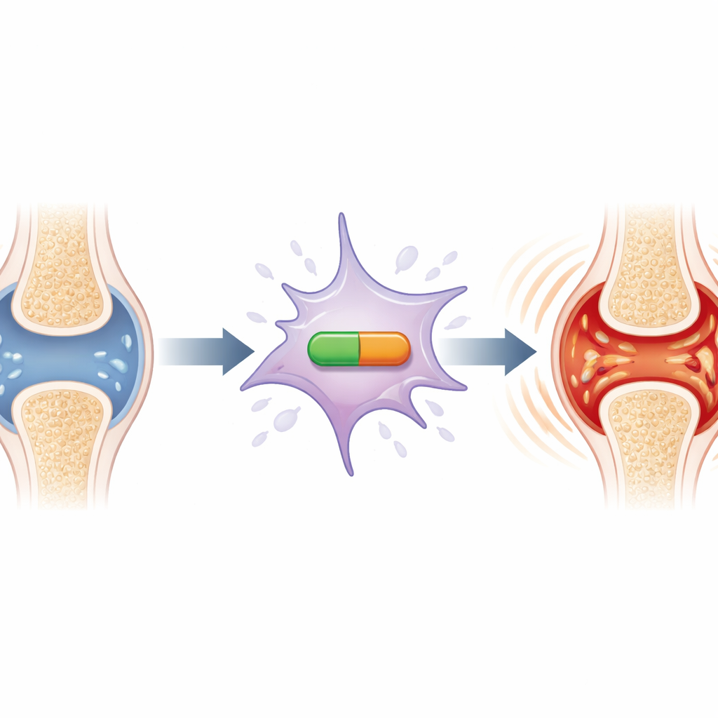
Why joint lining cells matter in arthritis
Rheumatoid arthritis is best known for painful, swollen joints, but behind the scenes a specific type of joint-lining cell helps keep the fire of inflammation burning. This study looks at why those cells, called fibroblast-like synoviocytes, behave abnormally in rheumatoid arthritis compared with osteoarthritis. The researchers focused on a little-known molecular “brake” called NUB1 that normally helps calm inflammatory signals. They discovered that in rheumatoid joints this brake is not properly turned on, and that the reason lies not in damaged DNA, but in the way the DNA is chemically packaged and regulated—its epigenetic landscape. Understanding this hidden layer of control could open the door to new treatments that cool inflammation without broadly suppressing the immune system.
A missing brake in the joint lining
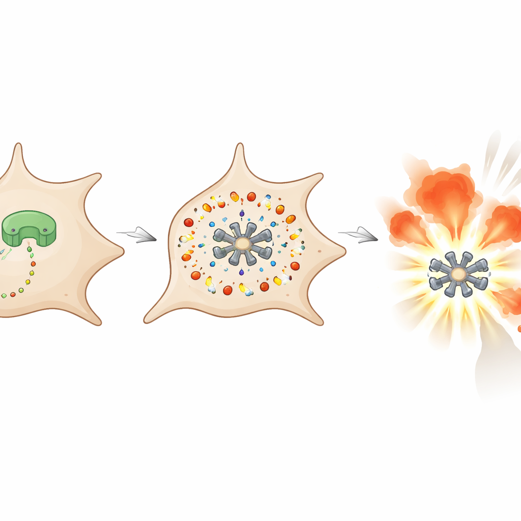
The inner lining of joints is made of a thin layer of cells that nourish cartilage and produce lubrication fluid. In rheumatoid arthritis, this layer thickens and fills with aggressive cells that churn out inflammatory molecules like interleukin-6 (IL-6). Earlier work showed that when these cells are stimulated by an inflammatory signal called IL-1, healthy or osteoarthritis cells can boost production of NUB1, a protein that acts as a brake on a pathway known as neddylation. Neddylation normally helps tag proteins for destruction and can switch on the master inflammatory regulator NF-κB. In rheumatoid cells, however, this inducible NUB1 response is blunted, allowing neddylation and NF-κB to run too hot and drive excess IL-6 production.
Seeing the imbalance inside real joints
To find out whether this imbalance actually occurs in patients’ joints, the team examined synovial tissue from people with rheumatoid arthritis and from those with osteoarthritis, a mostly wear-and-tear condition. Using antibody-based staining, they mapped where NUB1, the neddylation marker NEDD8, IL-6, and the NF-κB subunit p65 were located. In rheumatoid tissue, especially in the thin lining layer where fibroblast-like synoviocytes sit, NUB1 was noticeably lower, while NEDD8 and IL-6 were higher than in osteoarthritis tissue. Areas with low NUB1 showed strong nuclear localization of p65, indicating active NF-κB signaling. These spatial patterns support the idea that when the NUB1 brake is weak, neddylation and inflammatory signaling are stronger in the rheumatoid joint lining.
Ruling out simple signaling problems
The researchers then asked why rheumatoid fibroblast-like synoviocytes fail to ramp up NUB1 when exposed to IL-1. They compared baseline and stimulated NUB1 levels in cells from rheumatoid and osteoarthritis patients and confirmed that the basic, resting levels were similar, but the IL-1–driven increase was much smaller in rheumatoid cells at both the RNA and protein level. A series of tests ruled out common explanations. Blocking major signaling pathways known as MAP kinases did not normalize NUB1 induction, even though it reduced IL-6 as expected. The stability of NUB1 RNA over time was similar in both diseases, indicating the message was not being degraded faster in rheumatoid cells. A long non-coding RNA previously linked to NUB1, called SNHG12, and the activity of a core NUB1 promoter fragment also failed to account for the defect.
Epigenetic switches behind stubborn inflammation
With direct signaling and RNA handling largely excluded, the team turned to epigenetics—the chemical marks on DNA and histone proteins that help decide which genes turn on or off. Previous mapping had suggested that marks near the NUB1 gene differed in rheumatoid versus osteoarthritis fibroblast-like synoviocytes. In the new experiments, the researchers treated cells with drugs that broadly demethylate DNA, reduce a repressive histone mark, or block histone deacetylases, which generally makes nearby genes easier to activate. All three types of epigenetic drugs reduced or even erased the gap in IL-1–induced NUB1 between rheumatoid and osteoarthritis cells, without shutting down other IL-1 responses. This strongly suggests that abnormal epigenetic configuration in rheumatoid joint-lining cells keeps the NUB1 gene from turning on properly when inflammation strikes.
What this means for future treatment
In plain terms, this work shows that a protective brake on inflammation in joint-lining cells is not broken at the genetic level, but has effectively been “turned down” by epigenetic changes in rheumatoid arthritis. As a result, a chain of events—excessive neddylation, heightened NF-κB activity, and boosted IL-6 production—helps sustain chronic inflammation in the joint. By partially restoring NUB1 induction with drugs that remodel epigenetic marks, the study points toward new strategies that could calm inflammation by reprogramming resident joint cells rather than broadly suppressing the immune system. Such approaches might one day complement existing therapies and offer more targeted relief for people living with rheumatoid arthritis.
Citation: Ono, Y., Machado, C.R.L., Choi, E. et al. Altered fibroblast-like synoviocyte epigenetics is responsible for deficient NUB1 expression in rheumatoid arthritis. Sci Rep 16, 8128 (2026). https://doi.org/10.1038/s41598-026-38420-y
Keywords: rheumatoid arthritis, synovial fibroblasts, epigenetics, neddylation, inflammation