Clear Sky Science · en
Effect of a truncated mutant factor V on hemostatic function and embryonic development in mice
Why this matters for blood and baby development
Most of us only think about blood clotting when we get a cut, but the same system that stops a scraped knee from bleeding is also busy building and protecting blood vessels in the womb. This study looks at one key helper in that system, a protein called factor V, and asks a deceptively simple question: what happens to a developing mouse if this helper is badly damaged? The answer sheds light on a rare bleeding disorder in humans and on the hidden role clotting plays in making sure embryos survive and grow.
A hidden player in life and death
Factor V sits at a crossroads in the clotting process, helping the blood change from a liquid into a stable plug when a vessel is injured. People born with too little working factor V can suffer severe, sometimes life-threatening bleeding. Earlier work had shown that completely removing factor V in mice is usually fatal before or just after birth, but it was unclear exactly how this protein supports growing blood vessels. The authors set out to explore that link using a mouse strain created with gene-editing tools that unexpectedly produced a shortened, or truncated, version of factor V instead of the mild defect they had planned.
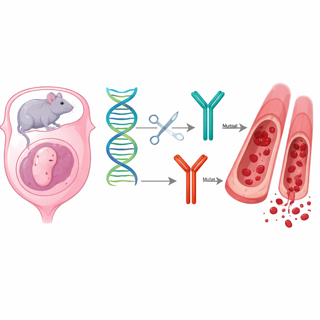
An accidental mutation with severe consequences
Using CRISPR gene editing, the team altered the factor V gene in mouse embryos. Alongside their intended mild change, a second variant appeared: a small deletion that shifted the reading frame and cut off the protein near its tail end. Mice carrying one normal and one mutant copy of the gene (heterozygotes) were born alive, but their blood tests showed that factor V activity dropped to about one-fifth of usual levels, and their clotting times were clearly prolonged. When two carriers were bred together, the expected mix of offspring was distorted. Far fewer animals carried two mutant copies, and most of those died around birth with widespread bleeding in the skin and organs or survived only a few weeks before dying without obvious external hemorrhage.
What the tissues reveal
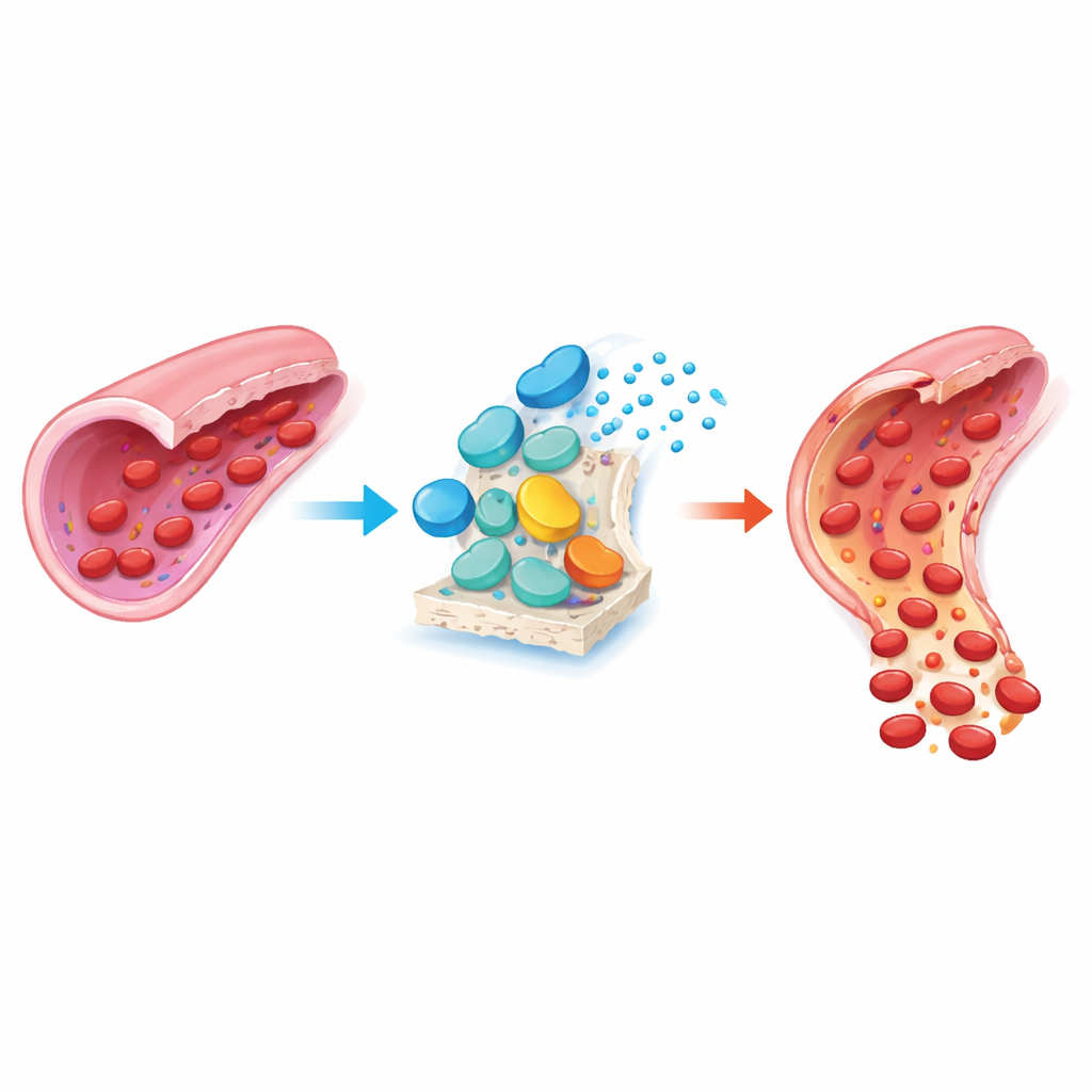
To understand what was going wrong, the researchers examined organs from affected and normal animals under the microscope and used staining techniques to track three key molecules: factor V itself, thrombin (the enzyme that actually makes clots), and a structural protein called alpha-smooth muscle actin that helps strengthen vessel walls. Newborn mice with two mutant copies showed congestion of blood vessels and small leaks in many tissues. In their brains and livers, signals for thrombin were much weaker than in normal pups, reflecting poor clotting activity. Signals for factor V were also reduced or absent outside the liver, suggesting that the truncated protein was made poorly, unstable, or not recognized by the stain. In the liver and heart, the smooth muscle layer around vessels stained faintly and looked thinner, implying that vessel walls themselves were underdeveloped and more fragile.
When and where factor V turns on
The team also measured activity of the factor V gene during different stages of mouse embryonic growth. They found that gene activity gradually rose from early stages to late gestation and then increased sharply in adult liver, confirming this organ as the main source. Looking across tissues, early embryos showed relatively high factor V gene activity in the yolk sac, a temporary, highly vascular organ that feeds the embryo before the placenta takes over. As development proceeded, the liver and a region that seeds future blood and vessel cells became the main sites of factor V production. These patterns fit with the idea that factor V supports early vessel formation in the yolk sac and later helps mature the growing circulatory system.
What this means for bleeding disorders
Taken together, the findings paint a picture in which a severely damaged factor V protein undermines both the ability of blood to clot and the proper building of blood vessel walls during development. In the mutant mice, reduced factor V leads to poor thrombin generation and weaker smooth muscle support around vessels, making them prone to leak and rupture. Many embryos likely die and are resorbed before birth, and those that are born face a high risk of fatal bleeding, especially in the brain. For people living with inherited factor V deficiency, these results help explain why some families experience miscarriages and why very low levels of the protein cause such severe disease. More broadly, the work underscores that the clotting system is not just an emergency repair crew, but an active partner in shaping and stabilizing the circulatory network that every embryo depends on.
Citation: Miguel-Batuecas, A., De Pablo-Moreno, J.A., Porras, N. et al. Effect of a truncated mutant factor V on hemostatic function and embryonic development in mice. Sci Rep 16, 8460 (2026). https://doi.org/10.1038/s41598-026-38387-w
Keywords: factor V deficiency, blood clotting, embryonic development, vascular biology, CRISPR mouse model