Clear Sky Science · en
Functional brain mapping of body size estimation using a 3D avatar
Why our sense of body size matters
Most of us assume we have a fairly accurate sense of our own shape and size, but this inner picture can be surprisingly distorted. For people with conditions like eating disorders or body dysmorphic disorder, those distortions can be severe and deeply distressing. This study asks a simple but powerful question: when we judge the size of our own body, what parts of the brain are doing the work, and how do they differ between people who are more or less accurate?
A digital mirror inside the MRI scanner
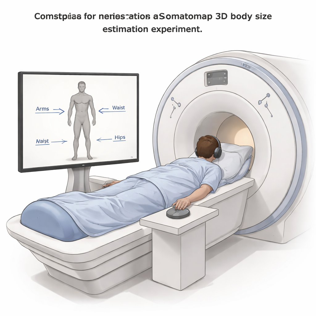
To explore this, researchers used a tool called Somatomap 3D, essentially an interactive digital mirror. Volunteers lay in an MRI scanner while viewing a three-dimensional, gender-matched avatar on a screen. Using a handheld trackball and sliders, they adjusted 26 separate body parts—from neck and shoulders to waist, hips, arms, and legs—until the avatar matched how they believed their own body looked. After scanning, the team carefully measured the same 26 body parts on each participant with tape measures, providing a physical reality check against each person’s internal image.
Comparing inner image to physical reality
By converting the avatar settings into centimetres, the scientists could compute, for every body part, how much each person over- or underestimated their size as a percentage of their actual measurements. Many body parts were slightly overestimated, but some areas around the midsection, such as waist and hips, tended to be underestimated. To capture each person’s overall pattern of errors, the team used a statistical approach that condenses complex, body-wide distortions into a few underlying “dimensions.” One of these dimensions reflected how consistently people misjudged the girth—the thickness—of body parts across the body.
Which brain regions light up when we reshape ourselves?
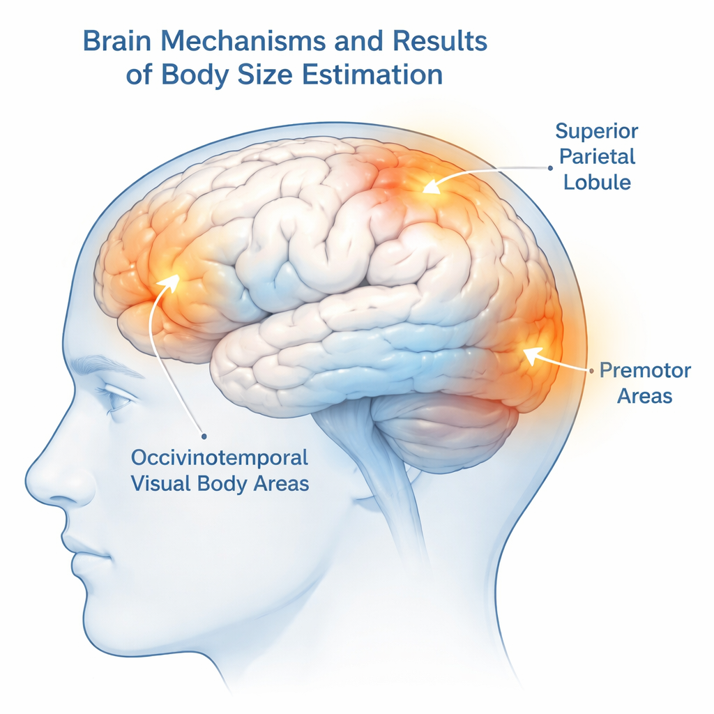
While participants were sculpting their avatars, the MRI scanner measured changes in blood flow, an indirect sign of local brain activity. Adjusting the avatar reliably engaged a network of regions already known to be involved in processing bodies and movement. Visual areas at the back of the brain that specialize in recognizing human bodies became active, as did premotor regions at the front of the brain that help plan and simulate movements. Crucially, a region near the top of the brain called the superior parietal lobule, which is involved in mapping space and integrating information about where our body is, also showed robust activity.
Linking brain activity to how accurate we are
The researchers then asked whether brain activity differed between people who were more versus less accurate in judging their own body size. Trial-by-trial accuracy—whether a single adjustment was slightly off or closer to reality—did not strongly predict moment-to-moment brain responses. However, when they looked at each person’s overall pattern of girth distortions across all body parts, one area stood out: the superior parietal lobule. People whose internal maps of body girth were more distorted showed a different level of engagement in this parietal region compared with those whose estimates were closer to their actual measurements. Other body-related visual and motor areas were active during the task but did not track these stable individual differences.
What this means for body image problems
To a layperson, the main message is that judging our own body size is not just a matter of looking in the mirror; it depends on a coordinated brain network that blends vision, spatial mapping, and mental simulation of our bodies. This study suggests that the superior parietal lobule may be especially important for maintaining an accurate internal map of how thick or thin our body parts are. Because problems with body size perception are central to disorders like anorexia nervosa and body dysmorphic disorder, pinpointing this region offers a concrete brain target for future research and, potentially, new treatments. The work also demonstrates that interactive 3D avatars can give scientists a more lifelike view of how we experience our own bodies from the inside out.
Citation: Peel, H.J., Diaz-Fong, J.P., Karsan, S. et al. Functional brain mapping of body size estimation using a 3D avatar. Sci Rep 16, 4750 (2026). https://doi.org/10.1038/s41598-026-38383-0
Keywords: body image, brain imaging, 3D avatar, body perception, eating disorders