Clear Sky Science · en
Single step nanosecond laser structuring for cost effective functional titanium surfaces with topography driven preosteoblast adhesion
Safer, Longer-Lasting Implants at Lower Cost
Millions of people rely on titanium implants to replace damaged teeth and bones, but not all implants fuse with the body equally well. A key challenge is getting bone cells to quickly and firmly attach to the metal surface so the implant becomes part of the skeleton. This study explores a simpler, cheaper laser treatment that sculpts the surface of titanium in a single step, creating tiny hills and valleys that encourage early bone cells to stick, spread, and grow—without needing the most expensive laser technology.
Why the Surface of an Implant Matters
When a titanium implant is placed in the body, bone does not simply glue itself to the metal. First, proteins from blood coat the surface, then bone-forming cells arrive, attach, and start building new tissue. How well this happens depends strongly on the surface texture and chemistry at scales too small to see with the naked eye. Earlier work suggested that the “best” implant surfaces must be highly oxidized and extremely water-loving, often achieved only with complex femtosecond lasers. Those systems are costly and hard to apply consistently to real-world implant shapes, which limits their widespread use in clinics.
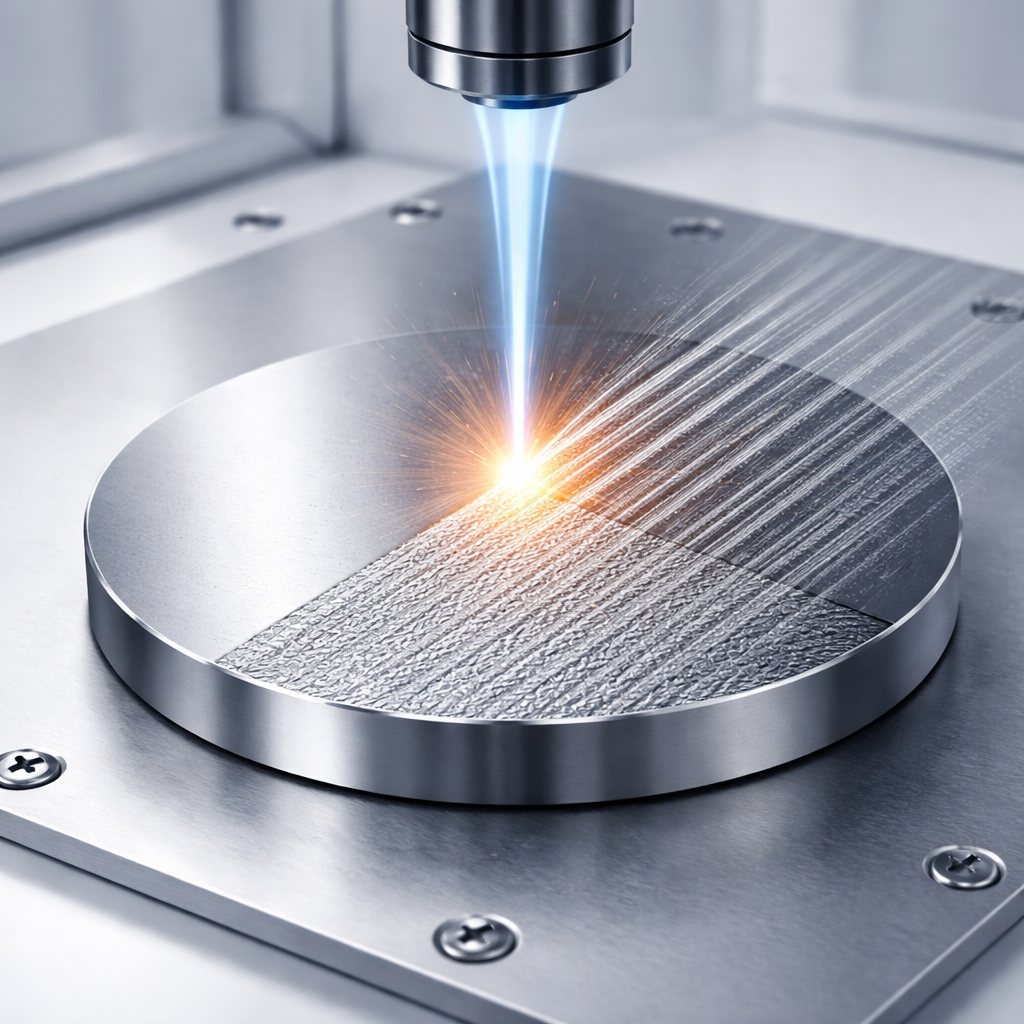
A One-Step Laser Carving Approach
The researchers used a more accessible nanosecond laser to pattern standard medical-grade titanium discs in a single processing step. By slightly changing the laser settings, they created two types of patterned surfaces, named P_0.4 and P_0.5, that differed mainly in the spacing of the laser tracks and resulting roughness. Powerful microscopes showed that both treatments produced uniform, rugged landscapes: broad grooves overlaid with spherical micro- and nano-sized bumps. Chemical analyses confirmed that the laser added only a modest amount of oxygen—forming a thin titanium oxide skin—while leaving the strong metal structure underneath unchanged. The treated surfaces were surprisingly water-repelling, with water droplets forming almost spherical beads.
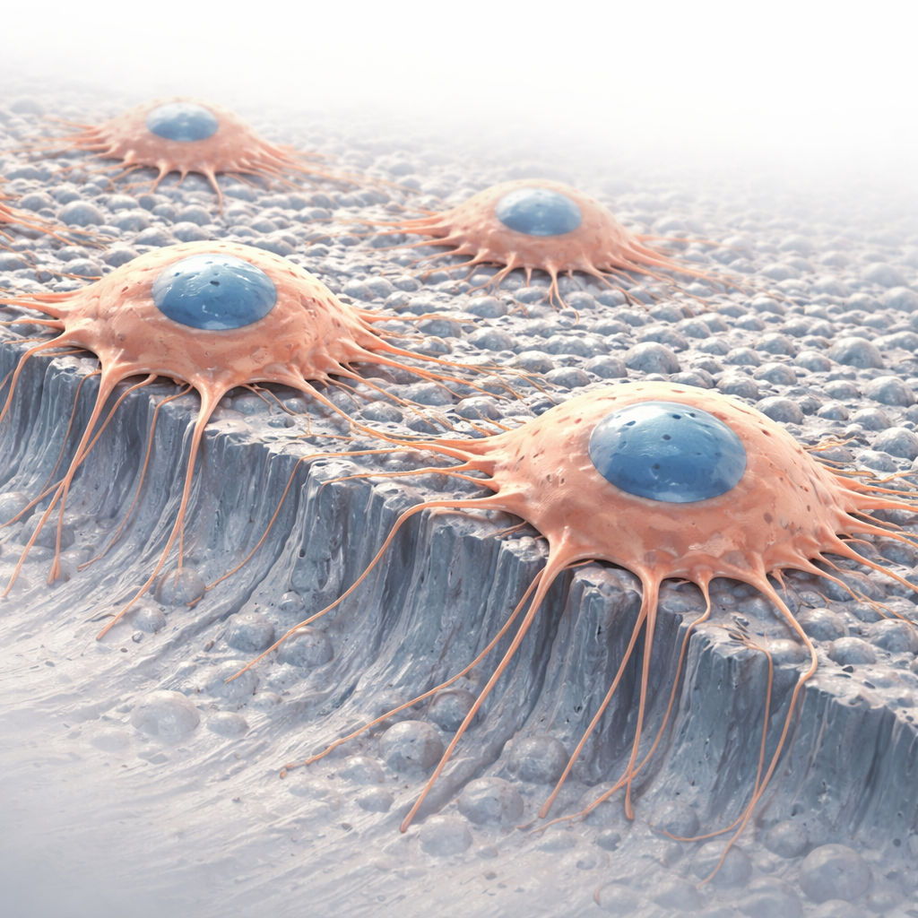
Testing How Cells Respond
To find out whether these unusual hydrophobic surfaces were friendly to bone, the team grew mouse preosteoblasts—precursor cells that develop into bone-building osteoblasts—directly on the laser-treated discs. They first checked for toxicity by measuring enzymes released from damaged cells and by fluorescent staining that distinguishes live from dead cells. Both tests showed that cells on P_0.4 and P_0.5 were as healthy as those on standard plastic used in cell culture labs. Over several days, the researchers then tracked how many cells accumulated and examined their shapes and internal scaffolding using confocal microscopy. On both laser-treated surfaces, the cell numbers steadily increased, and the cells spread out with well-developed support fibers, a hallmark of good attachment and growth.
Rethinking What Makes a Good Implant Surface
Perhaps the most striking result is that these moderately oxidized, strongly water-repelling titanium surfaces supported preosteoblast adhesion and proliferation just as well as more complex, highly oxidized, superhydrophilic surfaces reported in earlier work. The study also compared many published laser-treated surfaces with different roughness, oxygen content, and wettability. The pattern that emerges is that there is no single “magic” combination. Good cell responses can arise in two different regimes: smooth, highly water-attracting surfaces or rough, strongly water-repelling ones. In the latter case, the micro- and nano-scale texture seems to help proteins and cells find stable footholds, compensating for the lack of strong water attraction.
What This Means for Future Implants
For non-specialists, the takeaway is that making better implants is not just about exotic materials or the most powerful lasers. By carefully tuning surface texture with an affordable nanosecond laser, this work shows that it is possible to create titanium surfaces that bone cells like, without pushing oxidation and wettability to extremes. This single-step method could lower manufacturing costs, simplify quality control, and still provide a hospitable landscape for bone to grow onto the implant—potentially improving the comfort and longevity of joint replacements and dental implants for many patients.
Citation: Barylyak, A., Meskinis, S., Lazauskas, A. et al. Single step nanosecond laser structuring for cost effective functional titanium surfaces with topography driven preosteoblast adhesion. Sci Rep 16, 7104 (2026). https://doi.org/10.1038/s41598-026-38369-y
Keywords: titanium implants, laser surface texturing, bone cell adhesion, osseointegration, biomaterials