Clear Sky Science · en
Deep learning-based detection of retinal detachment with vitreous hemorrhage in ocular ultrasound images
Why this matters for saving sight
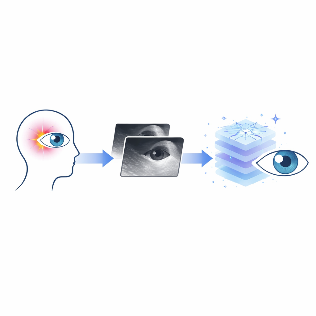
Retinal detachment is an eye emergency that can steal vision in a matter of hours or days. Doctors often rely on ultrasound scans when blood inside the eye blocks their view of the retina. But these grainy, echo-filled images can be hard to interpret, especially in busy emergency rooms or for less experienced clinicians. This study explores whether a modern form of artificial intelligence can quickly and reliably spot dangerous retinal detachments and related bleeding on ultrasound images, helping doctors protect patients’ sight.
Seeing through the haze in the eye
Two sight-threatening problems are at the heart of this work: retinal detachment, in which the light-sensing tissue peels away from the back of the eye, and vitreous hemorrhage, where blood leaks into the gel that fills the eye. When the eye is clear, doctors look directly at the retina to find trouble. But when dense blood clouds the view, they turn to ultrasound, which shows bright lines and speckled patterns bouncing off structures inside the eye. Unfortunately, the echoes from floating blood can look confusingly similar to the thin, sheet-like lines of a detached retina, leading to uncertainty at exactly the moment when rapid treatment matters most.
Teaching a computer to read eye scans
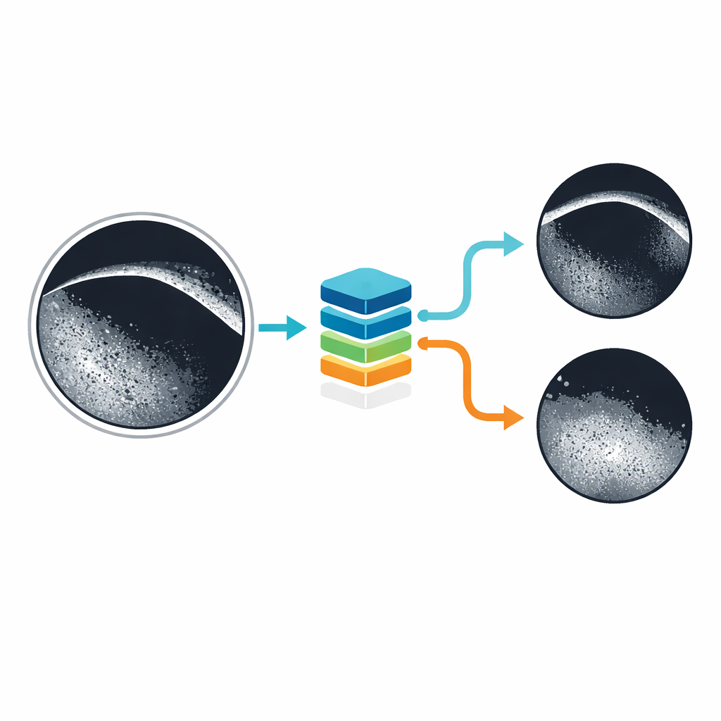
The researchers trained a deep-learning system, based on a real-time object-detection method known as YOLOv5, to pick out three possibilities on ultrasound images: retinal detachment alone, vitreous hemorrhage alone, or both together. They assembled 3,773 scan images taken over several years from patients already suspected of having these problems. Experienced eye specialists labeled each image and drew boxes around the areas showing disease, giving the computer examples of what to look for. The images were then split into separate sets for training, tuning, and final testing so that the system’s performance could be judged fairly on pictures it had never seen before.
Sharpening fuzzy pictures for the machine
Because ultrasound images are naturally blurry and speckled, the team tried several ways to make key structures stand out before feeding them to the AI. One method, called unsharp masking, subtly increases contrast around edges, making thread-like retinal detachments appear brighter and more distinct without adding obvious artifacts. They also experimented with thresholding and binarization—converting images into blocks of black and white based on brightness—to reduce the fog of scattered blood echoes while preserving the continuous lines that signal a detachment. In their main development process, they combined these enhancements with repeated training cycles and cross-validation, a strategy that helps avoid overfitting and improves reliability on new data.
How well the system performed
After several refinement rounds, the final model proved highly accurate when tested on 543 previously unseen images. It correctly recognized retinal detachment in 96.6% of cases, vitreous hemorrhage in 99.2%, and the especially tricky combination of both in 98.0%, yielding an overall accuracy near 98%. The researchers also compared different YOLO versions and found that, despite newer models performing well on general image benchmarks, YOLOv5 was better suited to this specific medical task and dataset. Additional experiments showed that while some preprocessing steps did not always raise average accuracy in isolation, they improved the clarity of key structures and appeared particularly helpful in the most visually confusing scans.
What this could mean for patients and doctors
For patients arriving in emergency departments with sudden vision loss, every minute counts. This study suggests that a carefully trained AI system could serve as a fast “second pair of eyes,” flagging retinal detachments and serious bleeding on ultrasound images with expert-level precision. The tool is not meant to replace ophthalmologists or the broader clinical examination, but rather to support them—especially when images are hard to interpret or specialists are not immediately available. Before such systems become routine, they will need testing across multiple hospitals, devices, and clinical workflows. Still, the results point toward a future where smart software helps doctors rescue vision more quickly and consistently when the retina is at risk.
Citation: Toyama, N., Hidaka, T., Tamura, H. et al. Deep learning-based detection of retinal detachment with vitreous hemorrhage in ocular ultrasound images. Sci Rep 16, 8398 (2026). https://doi.org/10.1038/s41598-026-38272-6
Keywords: retinal detachment, vitreous hemorrhage, ocular ultrasound, deep learning, medical imaging AI