Clear Sky Science · en
Enhancing diagnostic safety with low iodine, low radiation CTPA classification using deep learning
Safer Scans for a Dangerous Lung Clot
Pulmonary embolism is a sudden blockage in the blood vessels of the lungs that can be rapidly fatal if missed. Doctors rely on a special CT scan, called CT pulmonary angiography (CTPA), to spot these clots. But today’s most reliable scans often use relatively high doses of X‑ray radiation and iodine-based contrast dye, which can stress the kidneys and raise lifetime cancer risk. This study explores whether modern artificial intelligence (AI) can preserve the life‑saving accuracy of CTPA while using much less radiation and contrast dye, potentially making these scans safer for vulnerable patients.
Why Current Scans Come with a Trade‑Off
Standard CTPA produces crisp images of lung blood vessels by combining strong X‑ray beams with a generous dose of iodine contrast, which makes vessels glow on the scan. That clarity helps radiologists see small clots but comes at a price: repeated imaging can contribute to cumulative radiation exposure, and the contrast dye can harm patients with fragile kidneys or heart problems. When radiology teams try to reduce radiation or iodine, the images become grainy and dim, making subtle clots hard to distinguish from normal anatomy. Traditional computer algorithms, and even many deep‑learning tools, were built for full‑dose scans and tend to stumble when image quality drops.
A Two‑Step AI Helper for Low‑Dose Imaging
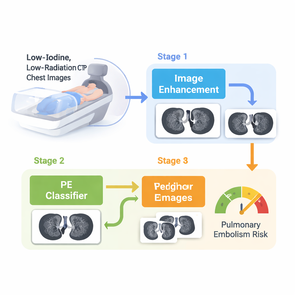
The authors designed a two‑stage AI framework tailored specifically for low‑iodine, low‑radiation CTPA. In the first step, an "image enhancement" network sharpens the blurry, noisy scans. It works by analyzing both the usual pixel patterns and their underlying frequency content—essentially separating fine edges, vessel outlines and subtle texture from background noise—then boosting the important details while suppressing clutter. In the second step, a "dual‑branch" classifier looks at both the original low‑dose image and its enhanced version side by side. One branch focuses on the overall structure of the chest, while the other zooms in on fine vessel details. The system then fuses these two perspectives with an attention mechanism that learns when to trust which branch most.
A New Real‑World Dataset and How It Was Tested
To make this approach clinically meaningful, the team assembled a new dataset of 191 adult patients scanned at Beijing Hospital using deliberately reduced radiation and only 30 milliliters of iodine contrast—substantially less than the 50–100 milliliters often used in standard protocols. Experienced radiologists labeled each case and, for a subset, painstakingly outlined clot‑containing slices. The researchers also created simulated low‑dose images from a large public dataset to pre‑train their models before fine‑tuning on the real low‑exposure scans. They then measured performance using standard diagnostic metrics such as sensitivity (how many true clots are found), specificity (how many false alarms are avoided) and the area under the ROC curve, a summary of overall accuracy.
Sharper Images and More Reliable Clot Detection
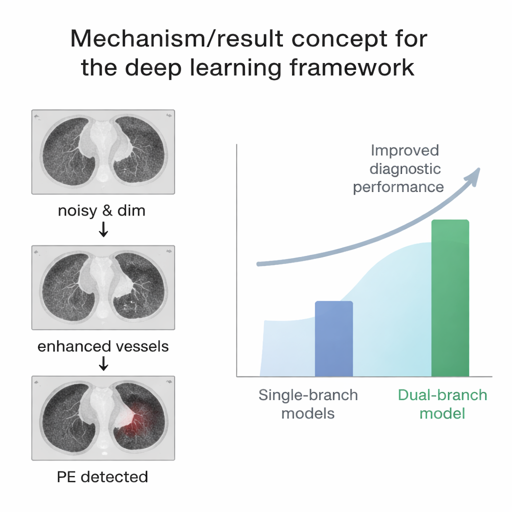
The enhancement network alone produced clearer vessel images than several well‑known super‑resolution methods, preserving fine structures while limiting artificial "hallucinated" details. Yet using only the enhanced scans for diagnosis did not outperform using the raw low‑dose images, because sharpening can sometimes exaggerate harmless patterns that mimic disease. The real advance came from the dual‑branch design: by combining the stability of the original images with the extra detail of the enhanced ones, the system achieved a high area under the ROC curve of 0.928, with balanced sensitivity and specificity. It also remained robust when extra noise was added, suggesting it can cope with the imperfect conditions of real‑world low‑dose imaging.
What This Could Mean for Patients
For patients, the key message is that AI may help make essential scans for pulmonary embolism safer without sacrificing reliability. The study shows that a carefully designed, task‑aware AI system can compensate for some of the quality loss that comes with lower radiation and less iodine contrast. That could be particularly valuable for people who need repeated imaging, or whose kidneys or overall health make standard contrast doses risky. While broader testing across multiple hospitals and scanner types is still needed, this work points toward a future where life‑saving clot detection can be achieved with gentler, more patient‑friendly CT protocols.
Citation: Hong, M., Gu, T., An, H. et al. Enhancing diagnostic safety with low iodine, low radiation CTPA classification using deep learning. Sci Rep 16, 7205 (2026). https://doi.org/10.1038/s41598-026-38223-1
Keywords: pulmonary embolism, low-dose CT, CT pulmonary angiography, medical imaging AI, contrast agent reduction