Clear Sky Science · en
Brain lipid profiles and oligodendrocyte gene expression show discordant responses to high-fat diet in Alzheimer’s disease mice
Why Fat and the Aging Brain Matter
Many of us worry that eating too much fat might damage our brains as we age, especially with headlines linking obesity to Alzheimer’s disease. This study in mice tackles a surprisingly simple question with a complicated answer: what actually happens inside the brain’s fatty insulation, called myelin, when Alzheimer’s disease and a high‑fat diet collide? By tracking both the brain’s lipid (fat) makeup and the activity of the cells that build myelin, the researchers uncover a mismatch that challenges easy assumptions about diet and dementia.
The Brain’s Fatty Wiring
The brain is one of the oiliest organs in the body. Much of this fat sits in myelin, the insulating sheath that wraps nerve fibers and lets signals travel quickly and reliably. Myelin is packed with specialized lipids such as cholesterol, galactosylceramides, sulfatides, and ethanolamine plasmalogens. These molecules help keep the sheath compact, stable, and able to support fast communication. Damage to myelin or its lipids has been linked to inherited white‑matter disorders and is increasingly suspected to play a role in Alzheimer’s disease, where memory and thinking gradually decline.
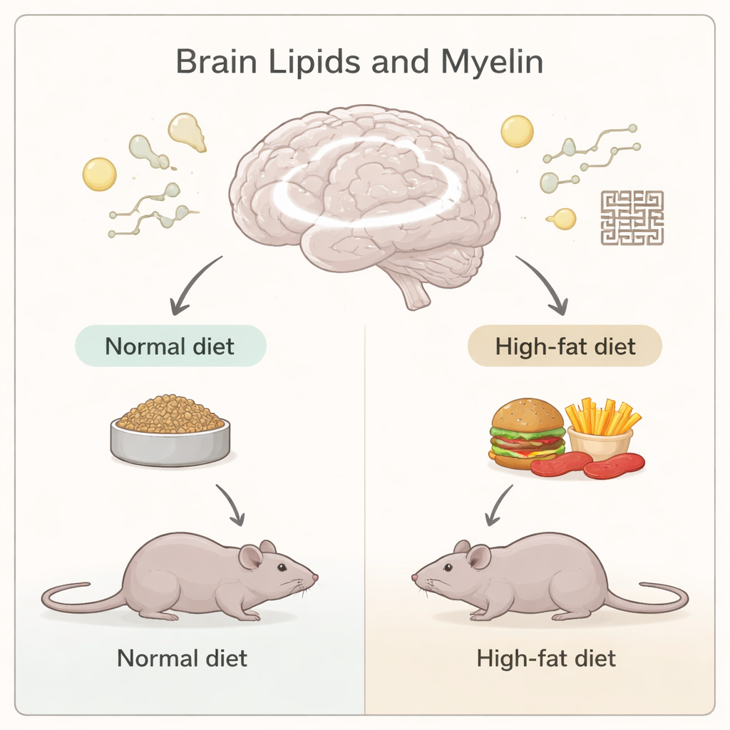
Alzheimer’s Mice on Different Diets
To probe how diet affects brain fats in Alzheimer’s, the team used a knock‑in mouse model that develops amyloid‑beta buildup and memory problems similar to early human disease. From two months of age, these Alzheimer’s mice and healthy control mice ate either a normal chow or a high‑fat diet that made them clearly obese. At seven to eight months, the scientists tested the animals’ spatial memory in a maze and then analyzed their brains. Using a broad survey of hundreds of lipid types in the cerebrum, they discovered that Alzheimer’s mice showed clear changes in many brain lipids, but the direction of change depended strongly on the diet.
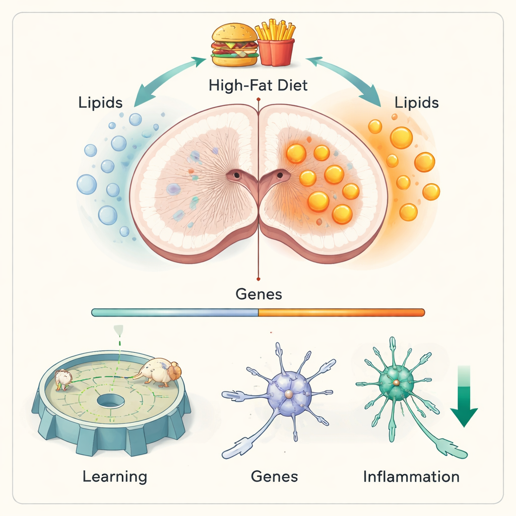
Fat Makeup Shifts, but the Builders’ Instructions Don’t
In Alzheimer’s mice on a normal diet, levels of several key myelin lipids—including certain galactosylceramides, sulfatides, and one plasmalogen—were reduced, while some cholesterol esters were higher. These shifts resemble patterns reported in human Alzheimer’s brains and hint that myelin structure may be subtly compromised. Strikingly, when the same Alzheimer’s mice were fed a high‑fat diet, these particular myelin lipids were no longer reduced; some plasmalogens were even increased. Yet when the researchers isolated oligodendrocytes, the cells that manufacture myelin, and examined their gene activity, the story was different. The expression of genes involved in myelin lipid pathways was altered by Alzheimer’s, but looked largely the same whether the mice ate normal chow or high fat. In other words, the blueprint inside the myelin‑making cells barely changed with diet, even though the actual lipid composition of the brain did.
Learning and Brain Inflammation Behave Unexpectedly
Given that midlife obesity raises dementia risk in people, one might expect a high‑fat diet to worsen memory and brain inflammation in these mice. Instead, the results were more nuanced. Alzheimer’s mice did show poorer spatial memory than healthy mice, as expected, but a high‑fat diet did not make this impairment worse. During repeated training in the Barnes maze, Alzheimer’s mice on the high‑fat diet actually learned the task faster than their counterparts on normal chow. Measures of brain immune activation told a similar story: markers of microglia and astrocytes—support cells that become reactive in Alzheimer’s—were elevated in Alzheimer’s mice, but high‑fat feeding did not further boost them. Notably, the astrocyte marker GFAP was lower in high‑fat Alzheimer’s mice, hinting at reduced astrocytic activation.
What This Means for Diet and Alzheimer’s
For non‑specialists, the central message is that brain fats and the genes that help make them do not always move in lockstep, and that a high‑fat diet in the context of existing Alzheimer’s‑like pathology did not simply make things worse. The mismatch between oligodendrocyte gene activity and diet‑dependent lipid changes suggests that additional layers of control—such as how enzymes are modified after they are made, how different brain cells share fats, and how lipids flow between the body and the brain—shape myelin health. While this work does not justify unrestricted high‑fat eating, it underscores that the impact of dietary fat on Alzheimer’s is complex, depending on age, disease stage, and the types of fats involved. Understanding these nuances could eventually help design more precise nutritional strategies to support the aging brain.
Citation: Kawade, N., Komine, O., Sobue, A. et al. Brain lipid profiles and oligodendrocyte gene expression show discordant responses to high-fat diet in Alzheimer’s disease mice. Sci Rep 16, 7224 (2026). https://doi.org/10.1038/s41598-026-38129-y
Keywords: Alzheimer’s disease, brain lipids, myelin, high-fat diet, oligodendrocytes