Clear Sky Science · en
AI-assisted radiographic analysis in detecting alveolar bone-loss severity and patterns
Why this matters for your next dental visit
Gum disease does more than make gums bleed—it quietly eats away the bone that holds teeth in place. Dentists try to spot this damage on dental X‑rays, but reading these images is difficult and time‑consuming, and small changes can be easy to miss. This study shows how artificial intelligence (AI) can help dentists measure bone loss around each tooth more quickly and consistently, opening the door to earlier treatment and better chances of saving teeth.
The hidden bone that keeps teeth in place
Each tooth is anchored by a support system of gum tissue, tiny ligaments, and jawbone. When long‑lasting gum infections are left untreated, this support slowly breaks down, leading to “alveolar bone loss,” the shrinkage of the bone that hugs the tooth roots. Worldwide, severe forms of this damage affect about one in five people over age 15 and are a major cause of tooth loss. On X‑rays, dentists gauge the severity of this loss by measuring the distance between a natural landmark on the tooth surface and the top edge of the surrounding bone, and they also look at the shape of that bone edge—whether it has sunk evenly (horizontal loss) or in a sharp, wedge‑like pattern (angular loss). Both the amount and the shape of bone loss matter for choosing the right treatment, including whether bone‑regrowth procedures are likely to work.
Why eyeballing X‑rays is not enough
Despite its importance, assessing bone loss on X‑rays is still largely done by hand and depends heavily on the dentist’s experience and level of fatigue. Two clinicians may give different readings on the same image, and busy clinics may struggle to examine every tooth surface in detail. Previous attempts to use AI in this area could often say whether bone loss was present, or roughly how severe it was, but they seldom gave precise, tooth‑by‑tooth measurements and usually did not capture both the severity and the pattern of loss at the same time. The authors of this paper set out to build a single automated system that could do both jobs—measure how much bone is gone and classify whether the loss is horizontal or angular—using the type of close‑up X‑rays dentists already take in everyday care.
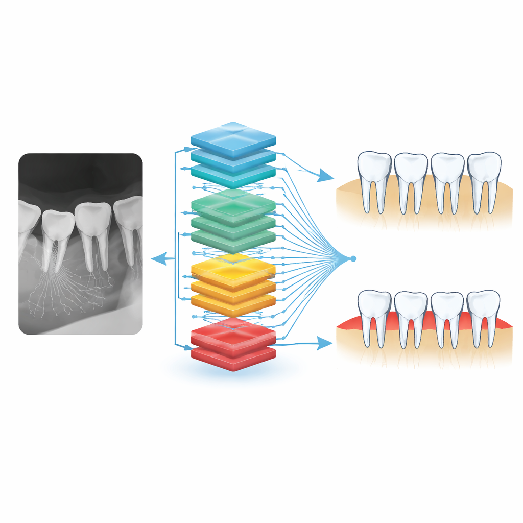
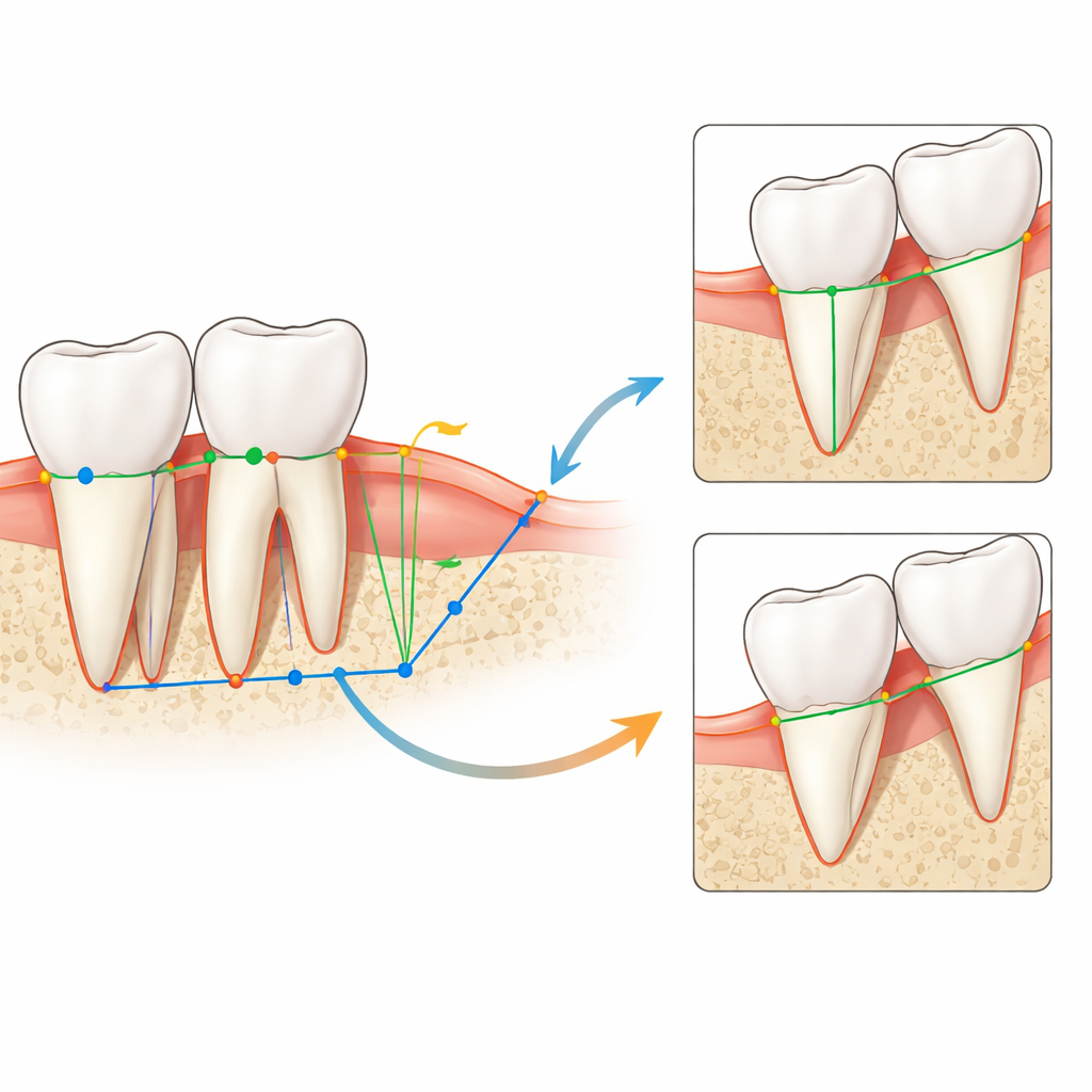
How the AI reads dental X‑rays
The team used a public collection of 1,000 carefully labeled intraoral periapical X‑rays, each showing several teeth in detail. First, an AI model was trained to find and draw rectangles around every tooth in an image. Within each tooth, a second family of models pinpointed three key locations: the enamel‑cement boundary near the gum line, the tip of the root, and the point where the visible bone edge crosses the tooth surface. By projecting these points onto a straight line and comparing their distances, the system turned pixel spacings into a percentage of bone loss for each tooth. A separate model traced the outlines of both teeth and bone edges, then converted those shapes into thin lines. At every site where bone loss was detected, the software compared the tilt of the tooth surface with the tilt of the bone line; shallow angles signaled angular defects, while steeper, more parallel lines indicated horizontal loss. This chain of steps allowed the AI to move from raw X‑rays to detailed measurements and pattern labels automatically.
How well the system performed
To check whether the AI’s measurements were trustworthy, the researchers compared them with expert annotations and with a dentist’s manual readings on a separate set of X‑rays. For bone‑loss severity, the agreement between the system and human experts was rated in the “good” range by standard reliability statistics, and the correlation with an experienced clinician’s estimates was strong. For bone‑loss patterns, the AI correctly distinguished horizontal from angular damage in about nine out of ten cases and showed moderate agreement with the expert’s decisions. The software also worked faster than a human reader: it could analyze a full X‑ray in roughly 25 seconds, compared with 1 to 4 minutes for a specialist, all while examining every tooth surface in a uniform way.
What this means for patients and dentists
The study suggests that AI can become a helpful second pair of eyes in the dental office, offering objective, repeatable measurements of how much bone has been lost and what kind of damage pattern is present. Dentists would still make the final call, but they could be alerted earlier to subtle changes, plan treatments with more confidence, and track whether therapy is slowing or stopping bone loss over time. Although the system still needs real‑world testing in everyday clinics, it points toward a future in which routine dental X‑rays double as precise, computer‑aided tools for preserving the bone that keeps our teeth in place.
Citation: Wimalasiri, C., Rathnayake, P., Wijerathne, S. et al. AI-assisted radiographic analysis in detecting alveolar bone-loss severity and patterns. Sci Rep 16, 7974 (2026). https://doi.org/10.1038/s41598-026-38061-1
Keywords: periodontitis, dental radiographs, artificial intelligence, bone loss, deep learning