Clear Sky Science · en
A hybrid approach for accurate skin lesion segmentation using LEDNet and Swin-UMamba
Why mapping moles matters
Skin cancer, including the dangerous form melanoma, often starts as a small, irregular spot on the skin. Doctors use special close-up photographs, called dermoscopic images, to study these spots, but carefully tracing the exact outline of each lesion by hand is slow and subjective. This study presents a new computer method that automatically draws highly accurate borders around skin lesions in such images, a step that can help earlier detection and more reliable monitoring of skin cancer.
From blurry borders to sharp outlines
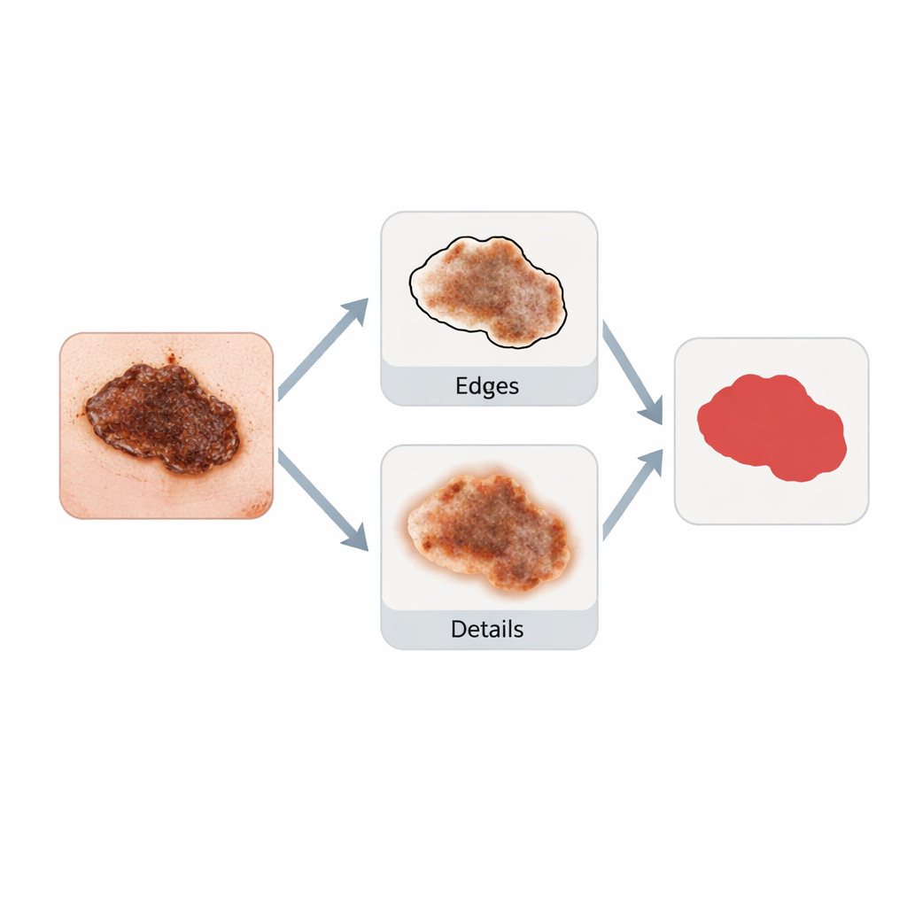
Traditional computer programs that analyze medical images are good at recognizing overall patterns but tend to “blur” the fine details where healthy skin meets suspicious tissue. For skin cancer, these edges are crucial: jagged or fuzzy borders can signal danger. Many existing systems either miss parts of a lesion or include too much surrounding skin, especially when the image is noisy, low contrast, or affected by hair and shadows. The authors argue that solving this problem requires a tool that sees both the big picture and the tiny, irregular details at the same time.
A two-part digital specialist
The researchers designed a hybrid system that combines two complementary components. The first, called LEDNet (Lesion Edge Detection Network), is dedicated to finding precise borders. It compares pairs of lesion images to highlight differences between the spot and nearby skin, then refines this information with an “edge guidance” module that produces a clean edge map—essentially, a thin outline of the lesion. The second component, Swin-UMamba, focuses on the overall structure of the image. It uses modern sequence-processing ideas, originally developed for long text and time series, to connect information from distant parts of the image and understand the lesion’s full shape and texture. Together, the edge-focused and context-focused modules reinforce one another, leading to cleaner, more trustworthy outlines.
Teaching the system with real-world skin images
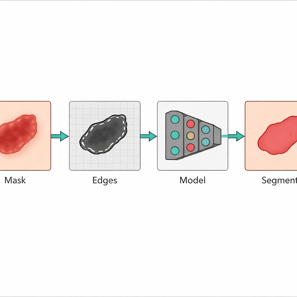
To see how well their approach works, the team tested it on three widely used collections of dermoscopic images: ISIC-2017, ISIC-2018, and Ph2. Each dataset includes skin photos along with expert-drawn masks marking where the lesion begins and ends. The researchers first generated simple edge maps from the existing masks using a classic technique called the Canny edge detector. These maps, along with the original images, were then fed into the hybrid model. Performance was measured using standard scores that compare the computer’s segmentation to the experts’ markings, including a Dice score, which approaches 1.0 when the match is almost perfect.
Results that rival expert tracing
Across all three datasets, the hybrid model outperformed well-known alternatives such as U-Net, attention-based networks, and other recent lightweight designs. On the ISIC-2017 and ISIC-2018 collections, the Dice scores were around 0.97, and on the high-quality Ph2 images they reached about 0.98, indicating a very close match to human-drawn borders. The method also showed high sensitivity (few missed lesion pixels), high specificity (few healthy pixels mislabeled as lesion), and strong overall accuracy. Visual heatmaps revealed that the system naturally focuses on the lesion boundary—the very area clinicians care about most—rather than being distracted by background artifacts.
Toward faster, more consistent skin checks
The authors conclude that their hybrid LEDNet–Swin-UMamba framework offers a powerful and efficient tool for automatically outlining skin lesions in dermoscopic images. By combining fine edge tracing with a global understanding of lesion shape, the method delivers segmentations that are both sharp and reliable, even for irregular or complex moles. While it will not replace dermatologists, such a system could become a valuable assistant—speeding up image review, reducing disagreement between experts, and helping ensure that suspicious changes in the skin are detected and monitored as early as possible.
Citation: Naeem, M.A., Yang, S., Saleem, M.A. et al. A hybrid approach for accurate skin lesion segmentation using LEDNet and Swin-UMamba. Sci Rep 16, 5415 (2026). https://doi.org/10.1038/s41598-026-38056-y
Keywords: skin cancer, melanoma, medical imaging, deep learning, lesion segmentation