Clear Sky Science · en
Prognostic value of the third thoracic vertebra skeletal muscle measurements in patients with digestive system malignancies: a comparative study with the third lumbar vertebra indices
Why Muscle Matters in Cancer Care
When people think about cancer, they often focus on tumors and treatments like surgery, chemotherapy, or radiation. But cancer also quietly reshapes the body itself, especially muscles. Losing too much muscle, a condition called muscle wasting, can leave patients weaker, less able to tolerate treatments, and more likely to have poor outcomes. This study asks a practical question with big implications: can a common chest scan, already used to diagnose and monitor many cancers, also double as a reliable tool to measure muscle health and help predict how long patients might live?
A New Way to Look Inside the Body
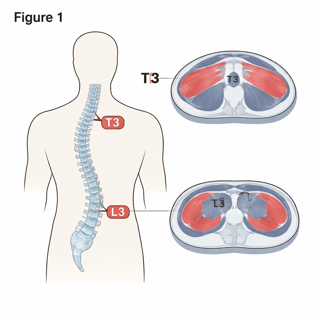
Doctors often measure muscle using a single slice from a computed tomography (CT) scan at the level of a bone in the lower back called the third lumbar vertebra, or L3. That spot has become the "gold standard" for estimating total body muscle. However, not every patient has an abdominal scan that shows L3. Many people with cancers in the chest, for example, have only chest CT scans. Those images do show another key landmark along the spine: the third thoracic vertebra, or T3, located behind the upper chest. The researchers wanted to know whether muscle measurements at T3 could serve as a stand-in for the usual L3 measurements.
Who Was Studied and How
The team reviewed records from 257 adults with cancers of the digestive system, such as colorectal, stomach, pancreatic, and liver cancers, treated at a single hospital in China between 2013 and 2018. Every patient had both chest and abdominal CT scans taken within one month of each other, along with standard information like age, height, weight, blood tests, and cancer stage. Using specialized software, the researchers carefully outlined the muscle on CT slices at T3 and L3 and calculated two numbers: the total muscle area at that level and an index that adjusts this area for a person’s height. They then followed patients over time to see who survived and who did not.
How Chest and Abdominal Measures Compare
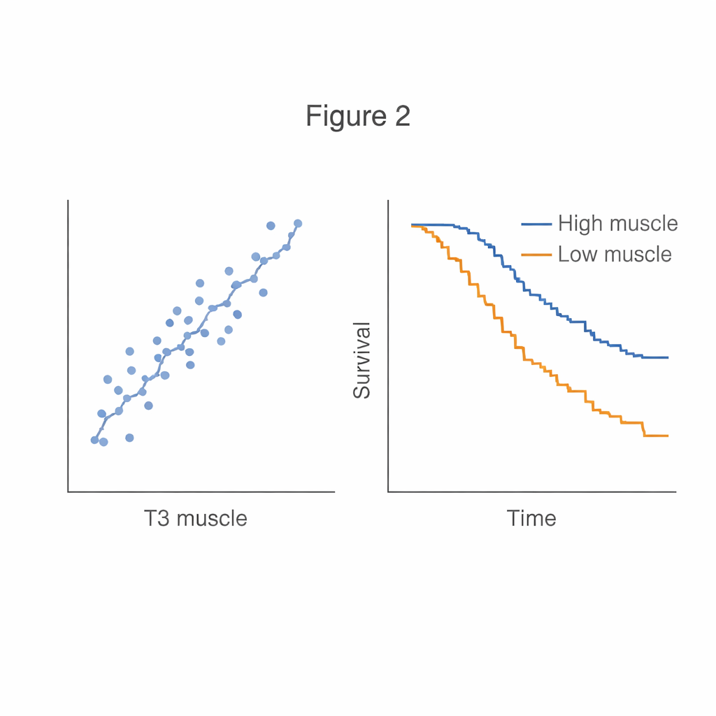
The central finding is that muscle measured at T3 closely tracks muscle measured at L3. Statistically, the two levels showed a strong correlation, meaning that patients with more muscle at T3 almost always had more muscle at L3, and those with less muscle at one level tended to have less at the other. This relationship held true across men and women, younger and older patients, and people with earlier or more advanced cancer. The researchers went a step further and built a mathematical formula that estimates L3 muscle area using T3 muscle area plus simple details such as age, sex, and body weight. This formula matched actual L3 values well, suggesting that in many cases a chest scan alone could provide nearly the same information as a full abdominal scan.
What Muscle Levels Say About Survival
Beyond the technical comparison, the study asked a more important question: do these muscle measurements tell us anything about a patient’s chances of survival? The answer was yes. Patients were divided into four groups based on how much muscle they had at T3 and at L3. Those in the lowest-muscle group were much more likely to die during follow-up than those with the most muscle, even after taking into account age, sex, cancer type, and stage. The risk rose sharply once muscle area or the muscle index dropped below certain cut-off values. In other words, thin muscles at either T3 or L3 signaled a higher risk, while fuller muscles were linked to better long-term outcomes.
What This Means for Patients
For people living with cancer, these findings suggest that images doctors already have on file can provide a useful snapshot of overall strength and resilience. A routine chest CT might quietly reveal who is at higher risk because of low muscle mass, even if they appear to have a normal body weight. Having this information could prompt earlier nutrition support, tailored exercise plans, or adjustments in treatment intensity. In simple terms, the study shows that looking at muscles in the upper chest can stand in for the usual measurements in the lower back, offering an easier way to spot vulnerable patients and, potentially, improve their care and survival.
Citation: He, Y., Li, Y., Zhao, Y. et al. Prognostic value of the third thoracic vertebra skeletal muscle measurements in patients with digestive system malignancies: a comparative study with the third lumbar vertebra indices. Sci Rep 16, 6749 (2026). https://doi.org/10.1038/s41598-026-37915-y
Keywords: cancer nutrition, muscle loss, CT imaging, sarcopenia, digestive system cancer