Clear Sky Science · en
Utilizing optical coherence tomography and machine learning to identify vision abnormalities in pediatric neurofibromatosis type 1 patients
Why this matters for children’s sight
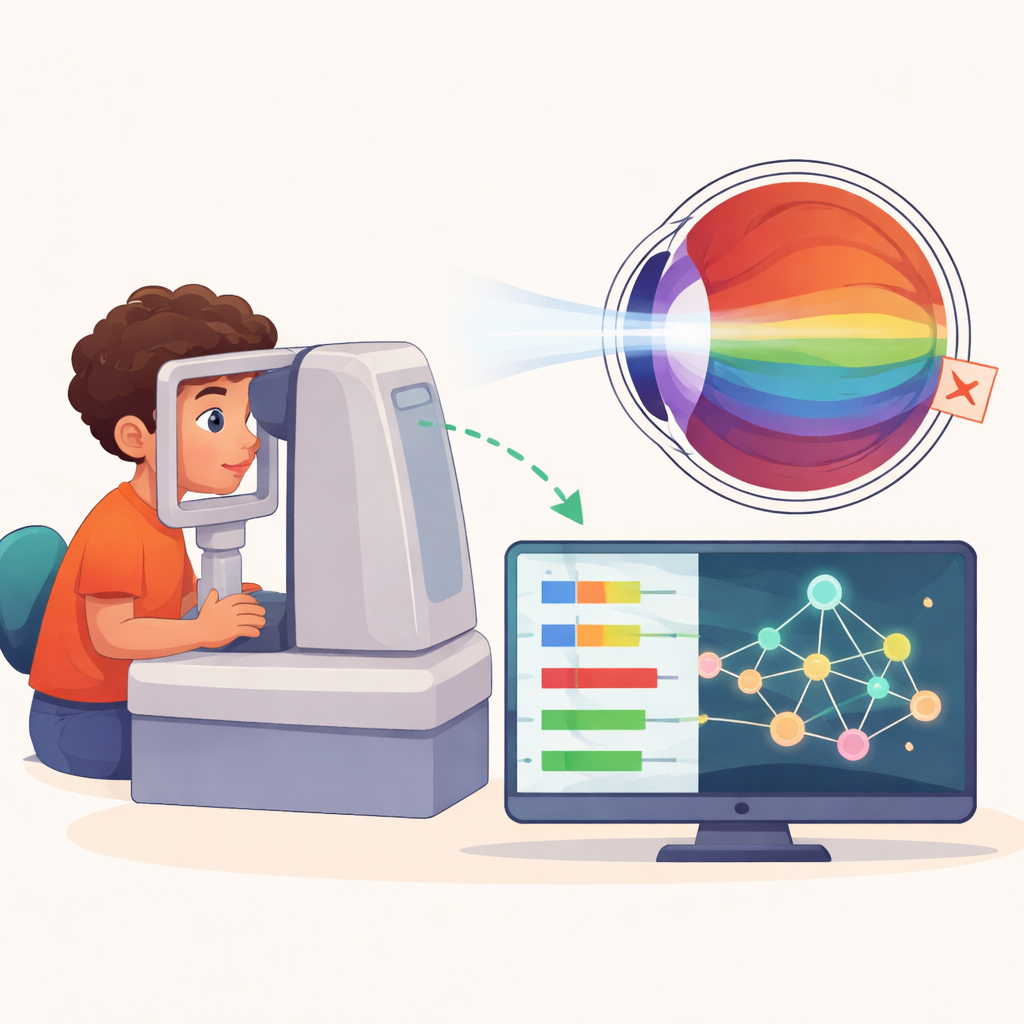
For children with a rare genetic condition called neurofibromatosis type 1, or NF‑1, losing vision can be a life‑changing complication. Doctors already use a non‑invasive eye scan called optical coherence tomography (OCT) to look at tiny structures at the back of the eye, but turning those images into clear warnings about which children are in trouble has been difficult. This study explores whether computer‑based pattern recognition, known as machine learning, can transform routine OCT measurements into an early‑warning system for vision problems in these young patients.
A genetic disorder with hidden eye risks
NF‑1 affects roughly one in 2,500 to 3,000 newborns and can cause a wide range of skin, nerve, and eye changes. One of the most serious threats is the development of tumors along the visual pathway, called optic pathway gliomas. About one in five children with NF‑1 will develop these tumors, often around age five, and some will suffer lasting loss of sharpness or field of vision. Because young children may not notice or report vision loss, doctors must rely on regular testing and brain scans to catch problems early—procedures that can be stressful, time‑consuming, and sometimes inconclusive.
A closer look at the eye’s wiring
OCT works a bit like ultrasound, but with light instead of sound, producing detailed cross‑section images of the retina and optic nerve. The research team focused on how thick or thin specific layers were, especially the retinal nerve fiber layer and the ganglion cell layers, which carry visual signals from the eye toward the brain. They collected 515 OCT exams from 168 children and teenagers aged 3 to 19, some with normal vision and others who showed reduced vision at one or more visits. Rather than analyze every pixel of every scan, the scientists used simple numeric summaries—overall thicknesses of key layers in the central retina (macula) and around the optic nerve—making the results easier to relate to real anatomy and to what clinicians already see in practice.
Training computers to spot early warning signs
The investigators then tried nine different machine learning approaches to see which could best tell normal from abnormal vision using only these thickness measurements. They paid special attention to avoiding over‑optimistic results by ensuring that data from the same child never appeared in both the training and testing sets. A model called a Balanced Random Forest—essentially an ensemble of decision trees tuned for unequal class sizes—emerged as the best fit for a screening tool. Using macular measurements alone, it correctly separated normal from abnormal vision with an area under the curve of 0.82 and detected about two‑thirds of children with vision problems, a level of sensitivity considered valuable when the priority is not to miss at‑risk patients.
From numbers to practical thresholds
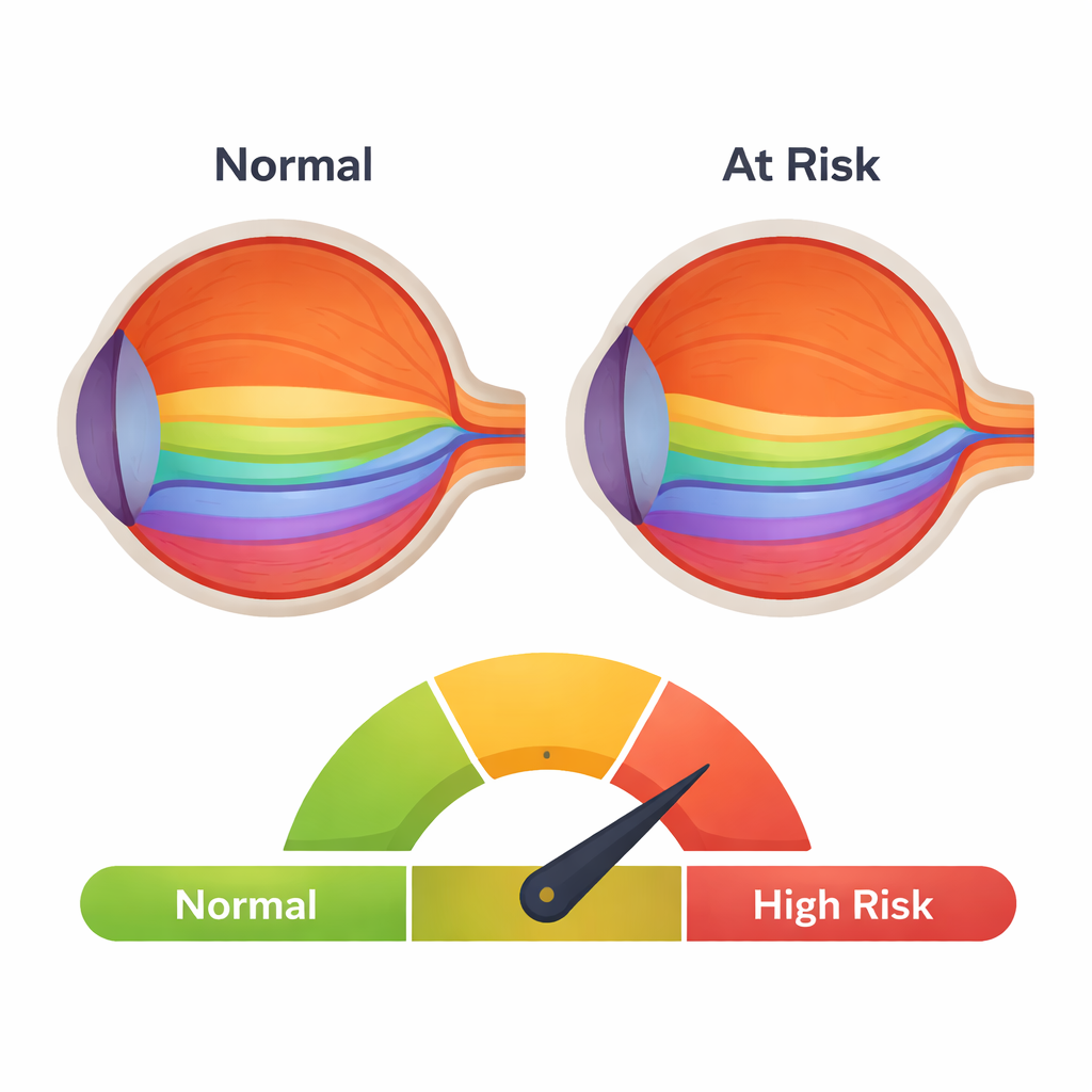
To keep the system understandable to clinicians, the team applied an explanation method that shows how much each feature pushes the model toward predicting normal or abnormal vision. This revealed that thinning of the retinal nerve fiber layer and the combined ganglion cell layers, in both the macula and the nerve region, was strongly linked with vision loss. The researchers went a step further, using these explanations to derive tentative cut‑off values—for example, a macular nerve fiber layer thinner than about 34 micrometers was associated with a much higher proportion of children with abnormal vision. They also tested how risk climbed as more layers crossed their cut‑offs: children exceeding three or more abnormal thresholds were far more likely to have vision problems than those who did not, suggesting that combining several subtle changes can sharpen risk estimates.
What this means for families and doctors
The study shows that simple measurements already available from standard OCT eye scans can be turned into a transparent, data‑driven tool to flag NF‑1 children who may be developing vision‑threatening damage. Rather than replacing doctors, such models could highlight which children need closer follow‑up, earlier treatment, or additional testing. The proposed thickness cut‑offs and “number of abnormal layers” rules are not yet ready to guide treatment on their own; they must be checked in larger, multi‑center studies. Still, this work suggests that combining precise eye imaging with explainable artificial intelligence could help protect sight in a vulnerable group of children by catching trouble before it becomes irreversible.
Citation: Cañada, C.F., Parcerisas, J.G., Bartomeu, J.P. et al. Utilizing optical coherence tomography and machine learning to identify vision abnormalities in pediatric neurofibromatosis type 1 patients. Sci Rep 16, 7237 (2026). https://doi.org/10.1038/s41598-026-37900-5
Keywords: neurofibromatosis type 1, pediatric vision, optical coherence tomography, machine learning, retinal nerve fiber layer