Clear Sky Science · en
3D CSFA-UNet: a unified attention-driven deep learning framework for accurate knee MRI segmentation and osteoarthritis severity classification
Why your knees – and this research – matter
Knee osteoarthritis is one of the main reasons people struggle with pain, stiffness, and even lose their independence as they age. Today, doctors usually judge its severity by eye on X-ray images, a process that can miss early damage and vary from one specialist to another. This study introduces a powerful artificial intelligence (AI) system designed to read both 3D MRI scans and standard knee X‑rays, automatically mapping joint structures and grading how bad the arthritis is. The aim is simple but important: faster, more reliable diagnoses that can guide treatment and surgery decisions with fewer guesswork and less manual effort.
Seeing more than the human eye
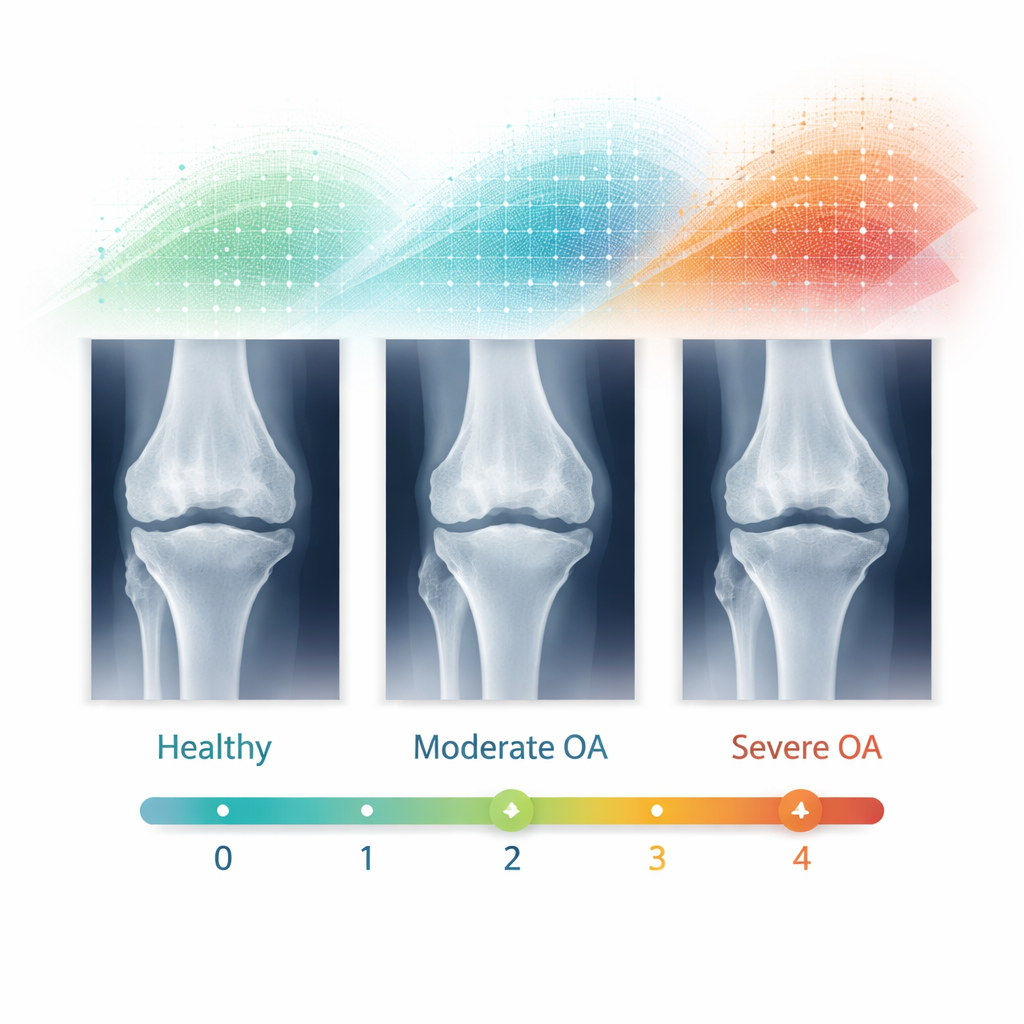
Traditional knee X-rays give a flat, two‑dimensional look at the joint. Doctors use the Kellgren–Lawrence scale, which runs from grade 0 (normal) to grade 4 (severe), to judge how worn the joint appears. But this approach often fails to spot the earliest changes, when cartilage has just started to thin and symptoms may be mild or vague. MRI scans tell a richer story: they show cartilage, meniscus, and other soft tissues in 3D, revealing subtle damage that X‑rays can’t see. The downside is that turning these scans into useful measurements usually requires painstaking, slice‑by‑slice tracing of structures by experts – far too time‑consuming to do for every patient in busy clinics.
A two‑lane AI highway for knee diagnosis
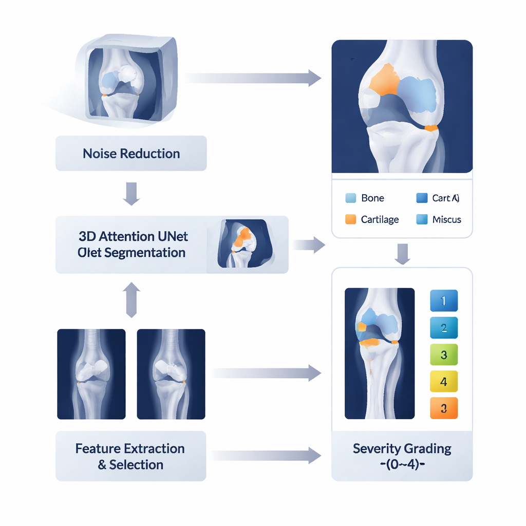
The authors built a unified AI framework with two coordinated lanes, each tailored to a different type of image. One lane takes in 3D MRI scans and first cleans them with a technique that sharpens tissue edges while reducing noise. These enhanced images go into a 3D "attention" U‑Net, a kind of neural network that not only looks at each tiny block of the image but also learns which regions and feature types matter most. It then produces detailed, color‑coded segmentations of the femur, tibia, and surrounding cartilage and meniscus. In parallel, a second lane analyzes ordinary knee X‑rays, extracting patterns at multiple scales – from fine edges to broader joint shapes – so that the system can relate what it sees on X‑ray to standard osteoarthritis grades.
From too many details to the most telling clues
Modern AI models can easily drown in their own information. To avoid this, the team introduces a feature‑selection step inspired by how desert scorpions hunt at night: they explore widely, then focus on the most promising vibrations in the sand. Here, a "Desert Scorpion" algorithm searches through thousands of numerical image descriptors and keeps only those that truly help distinguish one disease stage from another. These distilled features are then passed to a "spiking transformer" – a network that mimics how real nerve cells fire over time and how different parts of an image relate to each other. This classifier is further tuned by another nature‑inspired optimizer modeled on falcons that repeatedly adjust their flight paths as they close in on prey, searching the settings that make the model both accurate and stable.
Putting the system to the test
The researchers evaluated their framework on two public datasets: over 500 3D MRI scans with detailed labels of bones and cartilage, and 1,650 X‑rays graded from 0 to 4 for osteoarthritis severity. On MRI, the system’s segmentations of knee structures almost perfectly overlapped with expert‑drawn outlines, achieving a Dice score above 98 percent and very small distance errors measured in fractions of a millimeter. On X‑rays, it correctly identified the osteoarthritis grade more than 99 percent of the time, with very few missed cases or false alarms. When pitted against many existing methods – from classic convolutional networks to more recent multi‑task and transformer models – this combined pipeline was consistently more accurate, yet still efficient enough to be practical.
What this could mean for patients
In everyday terms, this study shows that a carefully engineered AI system can both "draw" the important pieces of the knee from 3D MRI and "judge" how worn the joint is from X‑rays with near‑expert precision. That opens the door to earlier, more objective detection of arthritis; better planning for total knee replacement; and large‑scale studies that track how the disease progresses or how treatments work, without requiring endless manual tracing by radiologists. While future work must confirm performance across more hospitals and imaging devices – ideally using paired MRI and X‑ray data from the same patients – this framework marks a major step toward computer‑assisted orthopaedic diagnostics that are fast, consistent, and easier to trust.
Citation: Moorthy, C., Shafeek, A., Gurunathan, V. et al. 3D CSFA-UNet: a unified attention-driven deep learning framework for accurate knee MRI segmentation and osteoarthritis severity classification. Sci Rep 16, 6878 (2026). https://doi.org/10.1038/s41598-026-37847-7
Keywords: knee osteoarthritis, medical imaging AI, knee MRI, X-ray grading, joint segmentation