Clear Sky Science · en
Three dimensional palatal morphology and dentoalveolar differences after extraction and non extraction treatment in class II malocclusion
Why the Roof of Your Mouth Matters in Braces
When people think about braces, they usually focus on straight teeth and a nice smile. But orthodontic treatment also reshapes the “roof” of the mouth—the palate—a space that helps guide the tongue, breathing, and how teeth fit together. This study asks a question many orthodontists face every day: when fixing a common bite problem called Class II malocclusion, does removing teeth (extraction) versus keeping them (non-extraction) change the three-dimensional shape of the palate in different ways?
Two Paths to Straight Teeth
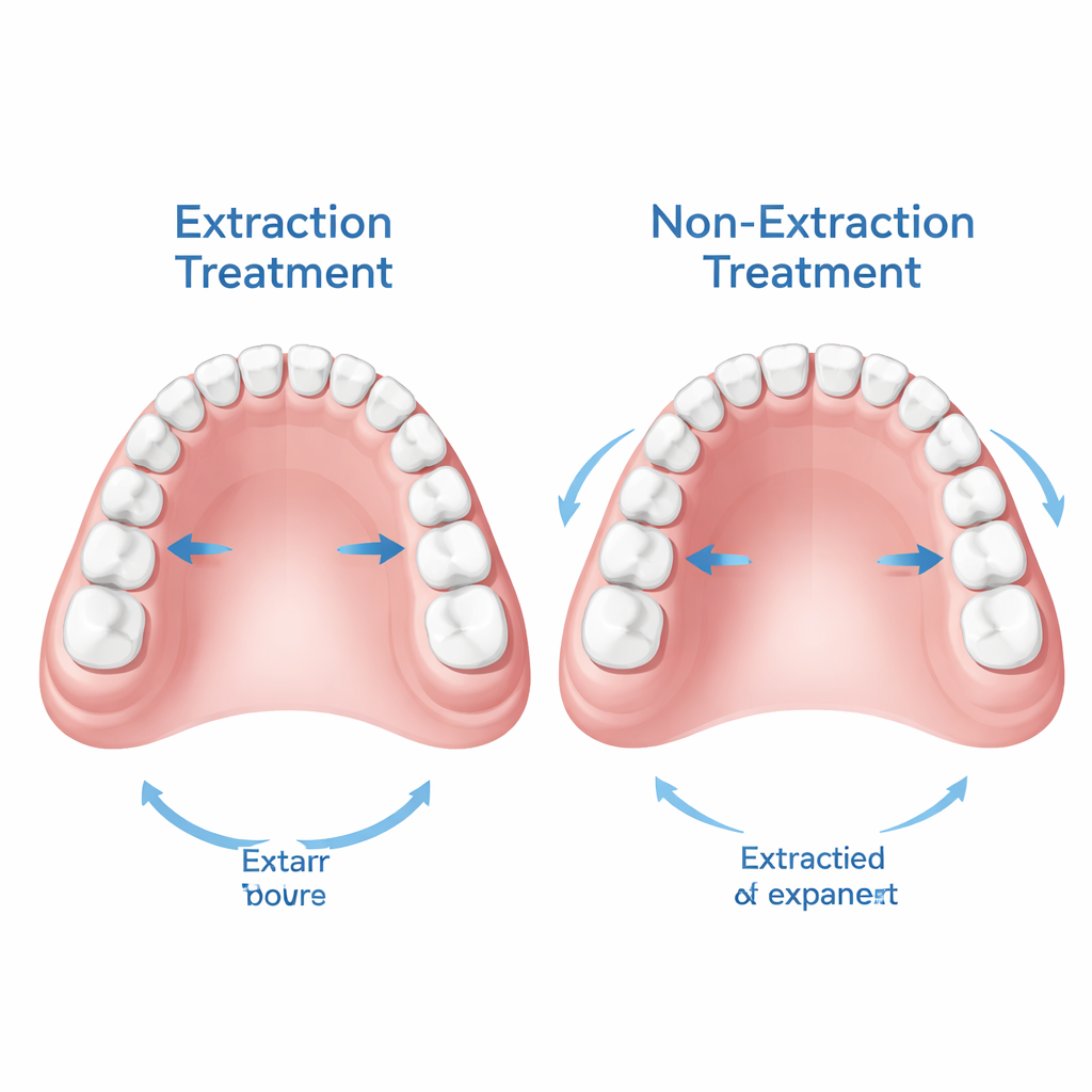
Class II malocclusion is a frequent bite problem where the upper teeth sit too far ahead of the lower teeth. In older teens and young adults, this is usually corrected with fixed braces rather than growth-guiding devices or jaw surgery. One major decision is whether to remove premolar teeth to create space or instead keep all teeth and make room by expanding and moving them. Supporters of extraction argue it helps manage crowding and protruding front teeth, while others prefer to widen and “develop” the dental arches without removing teeth. Until recently, most research looked at teeth and bones in two dimensions, giving little insight into how these different strategies reshape the palate in three dimensions.
Scanning the Roof of the Mouth in 3D
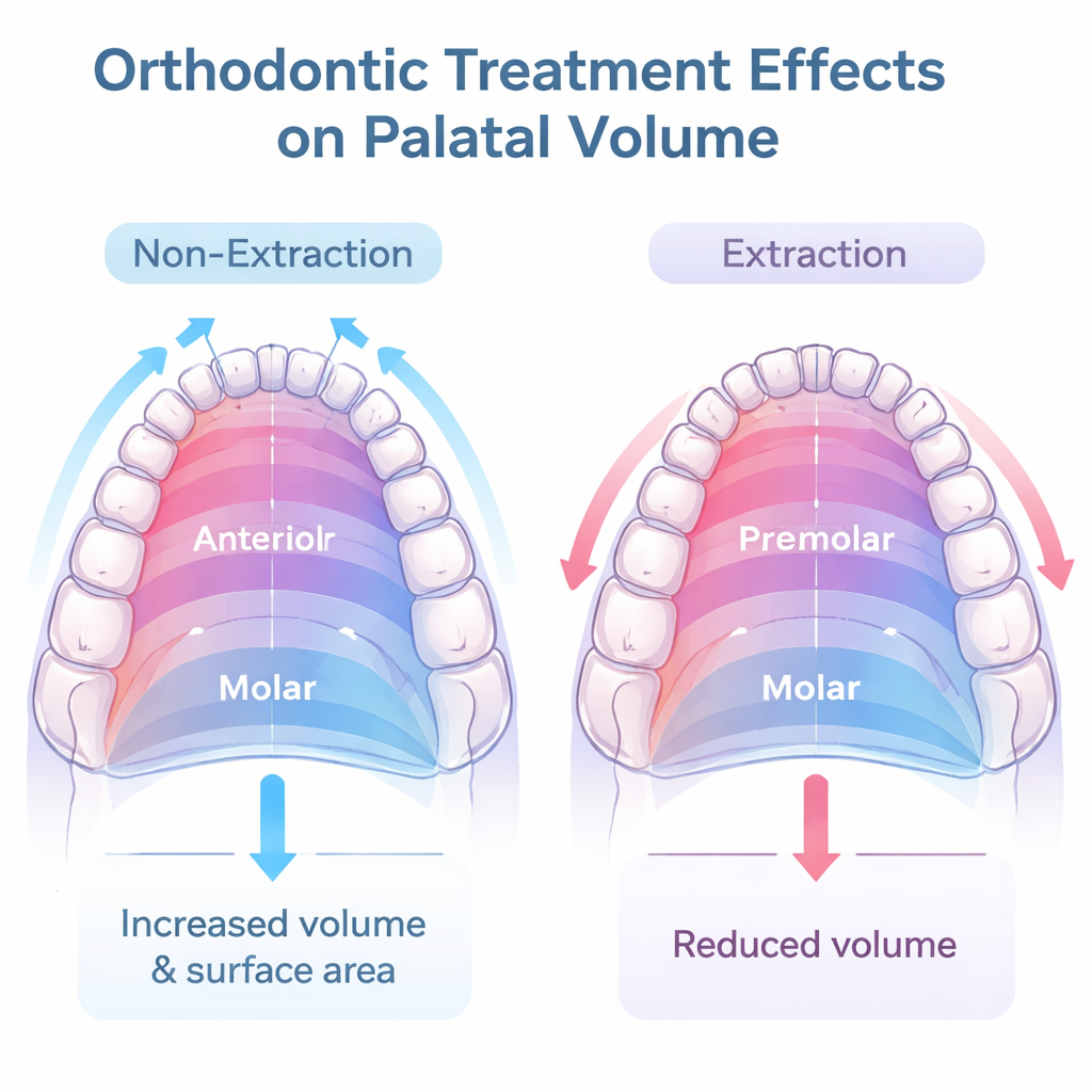
The researchers studied 69 post-pubertal patients: some with Class II malocclusion (the main treatment group) and others with Class I bite (a more regular control group). Each group was split into extraction and non-extraction subgroups. Before and after treatment, the team scanned plaster models of the upper jaw and used specialized software to measure palatal volume (how much space there is) and surface area (how large the inner surface is). They divided the palate into front, premolar, and molar regions and combined these into total volume and total surface area. At the same time, they analyzed head X-rays to track how the front and back teeth moved in different directions. All patients were treated with standard fixed braces, without special anchorage devices or expanders, so that differences mainly reflected whether teeth had been removed or not.
How Tooth Movement Reshapes the Palate
The overall jaw bones changed very little during treatment, but the teeth and palate did. In non-extraction cases, the upper front teeth tended to move forward, and the arches were often developed and slightly expanded. In extraction cases, first premolars were removed and the remaining teeth were pulled back to close the spaces. Across all groups, the front part of the palate gained volume and surface area, reflecting relief of crowding and better alignment of front teeth and canines. However, when looking at the entire palate, a clear pattern emerged: keeping all teeth (non-extraction) generally led to an increase in total palatal volume and surface area, while removing teeth (extraction) was linked to measurable reductions in these measures.
Direction of Tooth Movement Is the Key
To understand why these changes occurred, the authors used statistical models that linked tooth movement to changes in palatal shape. They found that forward or backward sliding of the upper front teeth and first molars—movement along the length of the jaw—was the main driver of palatal remodeling. When front teeth moved forward, palatal surface and, to a lesser extent, volume tended to increase. When back teeth were pulled forward to help close extraction spaces, total palatal volume and surface area tended to shrink. Vertical movements and angular tilting of teeth mattered much less once these forward–backward shifts were taken into account, suggesting that how orthodontists manage space along the dental arch is central to how the palate adapts.
What This Means for Patients and Clinicians
This study shows that the palate is not a static backdrop but a structure that reshapes itself alongside tooth movements. In older teens and young adults with Class II malocclusion, choosing extraction versus non-extraction treatment leads to different three-dimensional patterns of palatal change: non-extraction tends to enlarge palatal volume and surface area, whereas extraction tends to compact them. The work does not directly test how these changes affect breathing, speech, or long-term stability, but it highlights that digital 3D models can help orthodontists visualize and measure how treatment alters the space inside the mouth. For patients, the message is that the decision to remove or keep teeth influences not only how the smile looks from the front, but also how much room there is on the roof of the mouth—an important consideration in personalized orthodontic planning.
Citation: Rübendiz, M., Altunal, E.K., Kadıoğlu, M.B. et al. Three dimensional palatal morphology and dentoalveolar differences after extraction and non extraction treatment in class II malocclusion. Sci Rep 16, 6728 (2026). https://doi.org/10.1038/s41598-026-37842-y
Keywords: orthodontic extractions, Class II malocclusion, palatal volume, 3D digital models, dental arch development