Clear Sky Science · en
Analysis of the effect of pressure force on the microstructure properties of pressure measuring films
Seeing Invisible Forces
Whenever two surfaces touch—your foot on the ground, a gear tooth on its partner, or a surgeon’s tool on bone—forces spread across a tiny contact area. We can’t see those pressures with the naked eye, yet they decide whether a joint wears out, a medical implant succeeds, or a machine fails. This article looks inside a popular tool for revealing those hidden forces: pressure‑measuring films that change color when squeezed. The authors ask a simple but long‑ignored question: what is really happening inside these films when they are pressed?
How Color‑Changing Films Measure Pressure
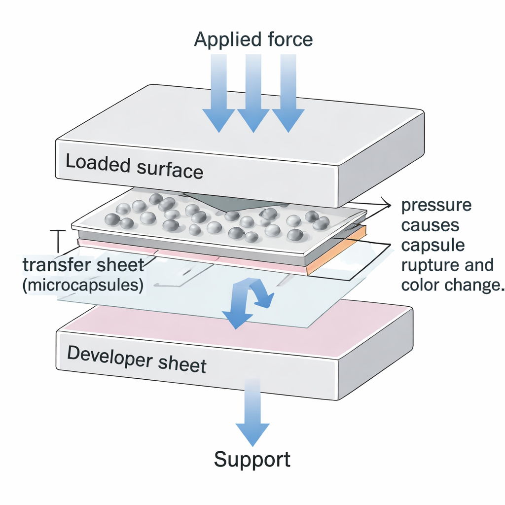
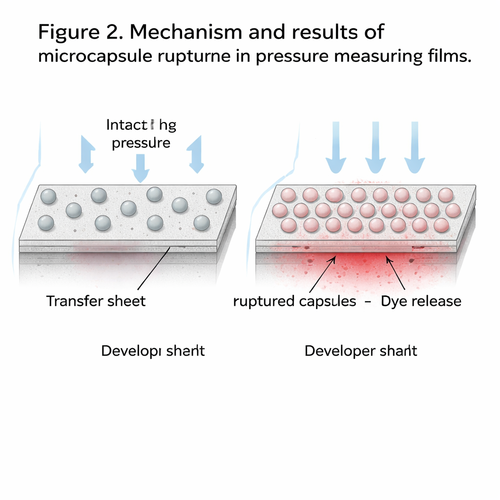
Commercial pressure films are thin plastic sheets that turn shades of pink or red where they are compressed. In the widely used two‑sheet version, one layer (the transfer sheet) carries countless microscopic capsules filled with a liquid dye. The second layer (the developer sheet) carries a special coating that reacts with that dye. When the sheets are sandwiched between two solid parts and pressed, some capsules burst and release dye onto the developer, creating a permanent color map of the pressure. Darker areas mean more capsules have ruptured, indicating higher pressure.
Looking Beneath the Surface
Previous work with these films focused on the colorful patterns they produce: how to calibrate color to pressure, or how to use the films in medicine, dentistry, and engineering. By contrast, this study zooms in on the film’s inner structure. Using a scanning electron microscope, the authors examined the transfer and developer sheets of a commercial two‑sheet system (SPF‑D from Sensor Products Inc.). They looked at areas that were never loaded, areas at the edge of the loaded zone, and regions directly under known forces. They also analyzed the chemical makeup of individual particles using X‑ray–based techniques.
The transfer sheet turned out to be a complex composite. The surface is crowded with smooth, spherical microcapsules ranging from about 1 to 40 micrometers in diameter—thousands of times smaller than a millimeter—mixed with tiny, bright mineral crystals. The capsules tend to form “grape‑like” clusters rather than a perfectly uniform layer. Chemical analysis showed that the capsules are mostly organic material containing the dye, while the bright particles are mainly calcium carbonate and other minerals that stiffen and stabilize the layer.
What Happens When You Press
To see how pressure damages the capsules, the researchers pressed small pieces of film between precision metal blocks under carefully controlled forces. They then counted intact and broken capsules in many microscopic regions. On average, each area the size of a grain of sand (about 640 by 480 micrometers) held roughly 900 capsules. About 2% were already damaged before any load—an important built‑in “background noise” for all measurements. As the applied force increased, the fraction of ruptured capsules rose steadily, but the way they broke stayed the same: capsules split in a characteristic, crater‑like fashion, often beginning with a fine crack across their diameter.
Interestingly, most of the action involves capsules of medium size, about 3 to 15 micrometers across. These mid‑sized capsules make up the majority of both intact and ruptured particles, meaning they largely control how much dye is released and how dark the imprint becomes. Very small or very large capsules are relatively rare. The clustering of capsules explains why the developer sheet does not color perfectly smoothly: local groups of densely packed capsules can release extra dye, creating small, darker spots even when the overall pressure is moderate.
The Other Half of the Sandwich
The developer sheet, which receives the dye, has its own important microstructure. It is a thin, brittle coating loaded with mineral pigments on a polyester base. Under the microscope, areas that experienced pressure show a network of fine cracks, like dried mud, while unloaded regions remain smooth. The same calcium‑rich particles found in the transfer sheet are even more abundant here, along with titanium and zinc compounds that likely affect color and opacity. This fragile, particulate layer helps trap and fix the dye, but its tendency to crack under load also limits how perfectly uniform the color can be.
Why This Matters for Real‑World Measurements
For users of pressure films in clinics, labs, and factories, these microscopic findings clarify why manufacturers quote an accuracy on the order of ±10–15%. Even before use, a small fraction of capsules are already broken, and the rest are clustered rather than evenly spread. Together with the brittle, cracked developer layer, these features introduce unavoidable variation into the color response. The study shows that despite this, the rupture process is highly consistent and statistically predictable: as pressure rises, more of the same types of capsules break in the same way. That insight strengthens computer models and calibration methods, helping engineers and clinicians interpret the colorful imprints more reliably and design better experiments, devices, and treatment plans based on what these seemingly simple films reveal.
Citation: Kalina, A., Ostachowski, P., Pytel, M. et al. Analysis of the effect of pressure force on the microstructure properties of pressure measuring films. Sci Rep 16, 7085 (2026). https://doi.org/10.1038/s41598-026-37837-9
Keywords: pressure-sensitive film, microcapsules, contact pressure mapping, materials microstructure, experimental mechanics