Clear Sky Science · en
Development and validation of a nomogram for predicting pulmonary embolism recurrence using muscle and fat parameters
Why body shape inside the chest matters
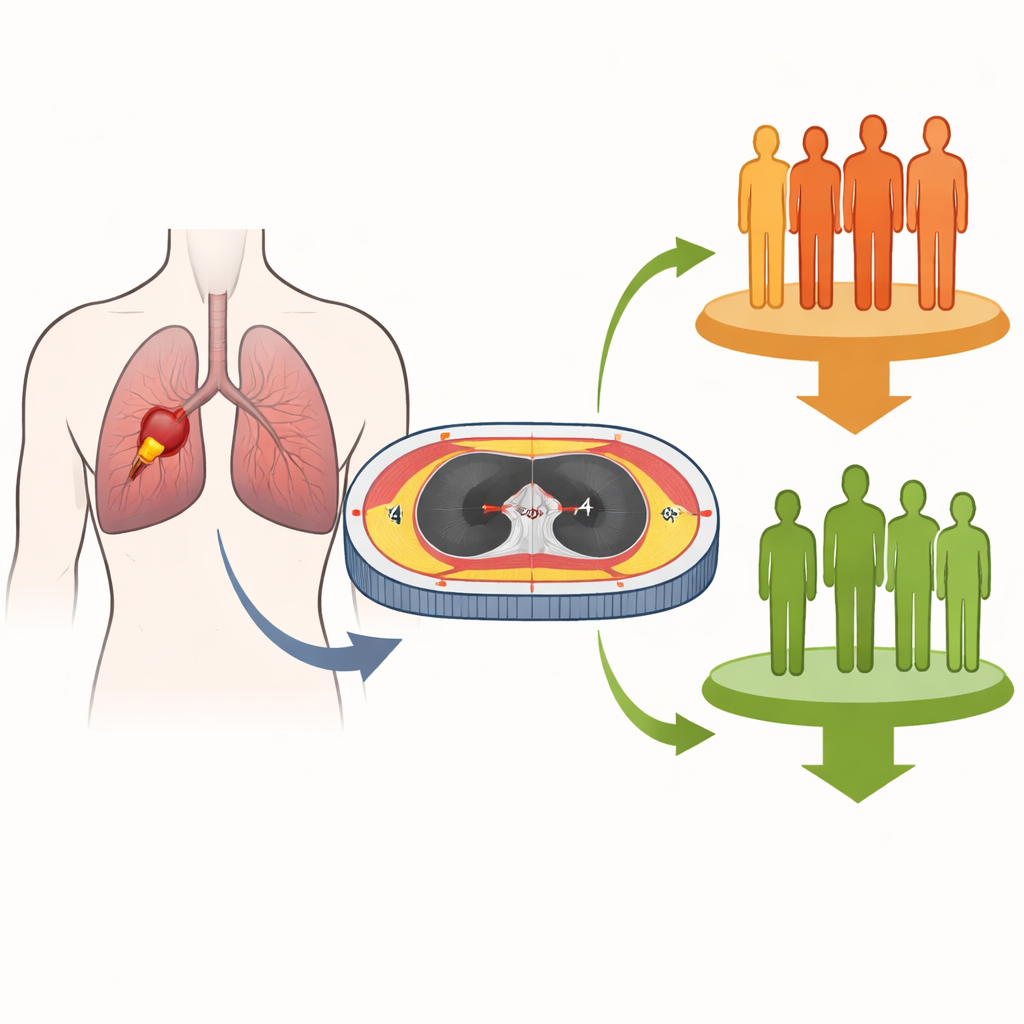
Pulmonary embolism, a blood clot that lodges in the arteries of the lungs, can be deadly not only the first time it occurs but also if it comes back. Doctors would like to know which patients are most likely to suffer a repeat event so they can tailor treatment and follow-up. This study explores a surprisingly simple data source that is already sitting in many patients’ medical records: the way muscle and fat are arranged inside the upper chest on routine CT scans. By turning these hidden body-shape clues into a prediction tool, the researchers hope to give clinicians a clearer picture of who is truly at risk.
Looking beyond the usual risk checklists
Current tools for judging clot risk in the lungs mostly rely on basic clinical information and blood tests: age, vital signs, lab values, and scoring systems such as the Wells or revised Geneva scores. These can flag patients at generally higher or lower risk, but they often miss important differences between individuals. At the same time, research in heart and lung disease has shown that the amount and quality of skeletal muscle and fat, especially around the chest, are closely tied to resilience, inflammation, and recovery. Yet these body composition measures have rarely been factored into predictions of whether a lung clot will return.
Turning CT scans into body composition maps
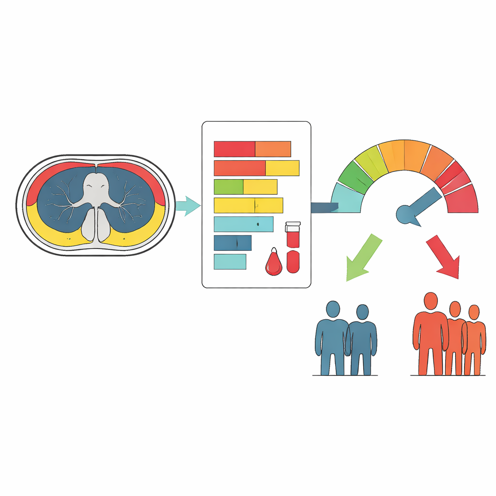
The team reviewed records from 184 adults who had a first diagnosis of pulmonary embolism between 2019 and 2023 and were followed for up to several years under standard blood-thinning treatment. All had undergone a specialized scan that combines nuclear imaging with low-dose CT. From a single slice of each CT scan at the level of the fourth upper back bone, the researchers used dedicated software to measure key features: the total area of chest muscles, the specific size and density of the pectoral muscles, and the amount and density of the fat just under the skin. These values reflect not only how much muscle or fat a person has, but also tissue quality, which can be altered by aging, poor fitness, or chronic illness.
Building a practical prediction tool
To see which factors best signaled the chance of a clot coming back, the investigators combined these imaging-based measures with standard clinical data such as body mass index, white blood cell count, and the presence of deep vein thrombosis in the legs. They used a statistical procedure that automatically filters out weaker variables and keeps only those that add meaningful information. Eight features emerged as the most useful, including chest muscle area and density, subcutaneous fat area and density, body mass index, white blood cell count, and leg vein clots. These were woven into a visual scoring chart called a nomogram, which allows clinicians to line up a patient’s values and read off an estimated probability of recurrence.
How well the model performed
The 184 patients were split into a larger group for building the model and a smaller group for testing it. When the nomogram was applied to the development group, it separated those who later had another embolism from those who did not with moderate accuracy; performance was somewhat lower, but still reasonable, in the test group. Just as important, the predicted risks closely matched what actually happened over time, and decision analyses suggested that using the tool would offer more benefit than simply treating everyone as high risk or low risk across a wide range of clinical scenarios. Notably, no single muscle-or-fat measure alone was decisive, but together they improved how finely risk could be graded.
What this means for patients
For people recovering from a lung blood clot, this study suggests that the hidden details of their chest muscles and fat, already captured on ordinary scans, may quietly signal how vulnerable they are to another event. By combining these imaging clues with routine clinical information in a single, easy-to-use chart, doctors could better identify who needs closer follow-up or longer protection with blood thinners, and who might safely avoid extra treatment. The authors caution that the model is an early step and must be tested in other hospitals and larger groups of patients. Still, it points toward a future where body composition becomes a routine part of personalizing care after pulmonary embolism.
Citation: Cao, J., Niu, S., Li, X. et al. Development and validation of a nomogram for predicting pulmonary embolism recurrence using muscle and fat parameters. Sci Rep 16, 8538 (2026). https://doi.org/10.1038/s41598-026-37833-z
Keywords: pulmonary embolism, body composition, computed tomography, risk prediction, recurrent thrombosis