Clear Sky Science · en
Altered histone modifications in Aedes aegypti midguts following Rift Valley fever virus exposure
Why mosquito genes matter for human health
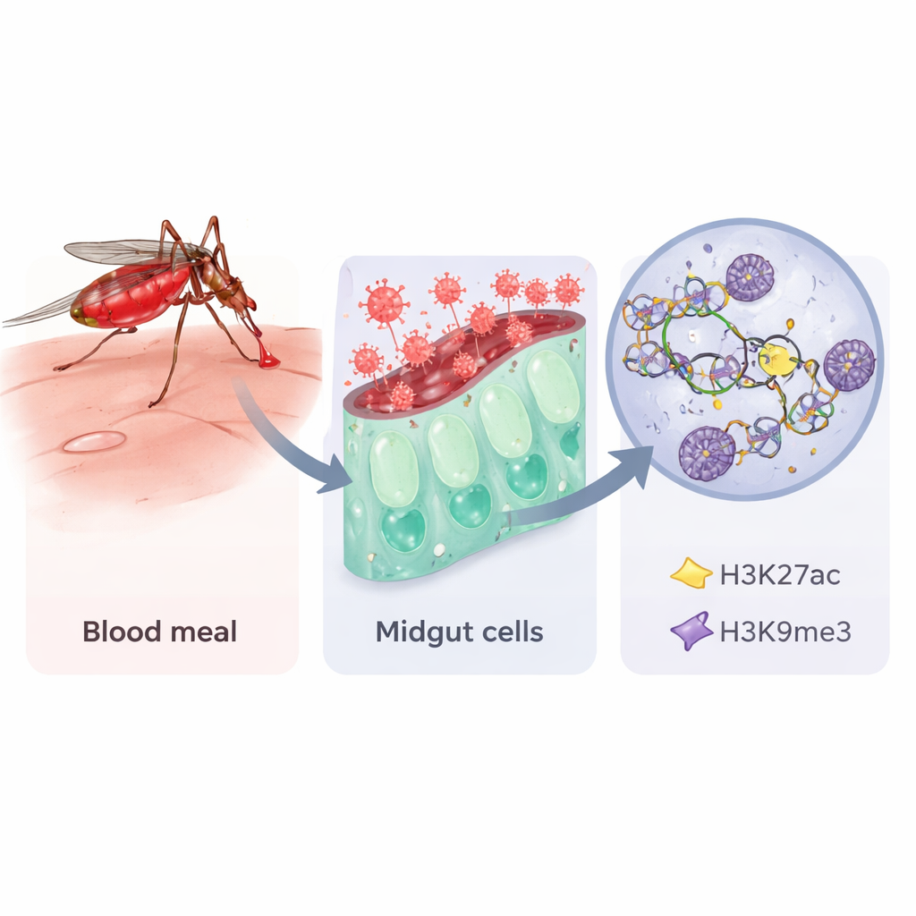
Rift Valley fever is a virus that sickens livestock and people in parts of Africa, and it is spread by mosquitoes. To keep outbreaks in check, scientists need to understand not only the virus itself, but also how mosquitoes respond when they swallow infected blood. This study looks inside the mosquito’s gut at an unusually fine level, asking how the virus nudges mosquito DNA packaging to turn critical genes on or off—changes that could influence whether the insect becomes a good or poor transmitter of disease.
Opening the book of mosquito DNA
Like people, mosquitoes do not change the letters of their DNA when they encounter an infection, but they can change how tightly that DNA is packed. The authors focused on two chemical tags on histone proteins, which act like spools for DNA. One tag, called H3K27ac, usually marks open, active stretches of DNA; the other, H3K9me3, is linked to shut‑down, silent regions. Using a sensitive method known as CUT&RUN, combined with RNA sequencing, the team examined these tags and gene activity in the midguts of Aedes aegypti mosquitoes after three kinds of meals: plain sugar, a normal blood meal, or blood containing a vaccine strain of Rift Valley fever virus. They sampled guts one, three and seven days after feeding to capture early, middle and later stages of infection.
How a simple blood meal rewires the gut
A blood meal alone, even without virus, caused sweeping changes in midgut gene activity. Thousands of genes shifted their activity one day after feeding, especially those involved in digesting proteins, handling energy, and building new cellular components. Many of these genes sat near regions marked by H3K27ac, consistent with the idea that blood triggers the gut to open up specific DNA neighborhoods needed for digestion and egg production. Over the next few days, as the blood was digested, the pattern evolved: energy‑producing machinery stayed active, and later, genes involved in organizing chromosomes and the cell cycle were affected. The sugar‑fed control mosquitoes, by contrast, showed more stable patterns, hinting that not taking a blood meal may lead to a different, possibly aging‑related, DNA landscape.
When virus meets midgut defenses
Adding Rift Valley fever virus to the blood changed the picture. Early on, one and three days after feeding, the midguts of virus‑exposed mosquitoes ramped up genes linked to immune defense and cell signaling, beyond the changes caused by blood alone. At the same time, the usual relationships between histone tags and nearby genes became more complicated. On day three in particular, many regions marked by the normally repressive H3K9me3 tag lost that mark, and hundreds of nearby genes became more active, including those involved in controlling other genes, relaying signals inside the cell, and managing cell shape and polarity. By seven days, when about half of the mosquitoes carried infectious virus, overall gene activity in exposed midguts dropped, immune‑related genes were dialed down, and activating H3K27ac marks were widely depleted compared with blood‑only controls.
Hints of viral tricks and mosquito defenses
By matching gene activity to nearby histone changes, the researchers identified a small set of genes whose behavior is especially suggestive. Some genes that help organize membrane structures or move materials inside cells gained activity while their local activating marks faded, making them candidates for helping the virus assemble or travel within the cell. Others, such as genes related to detoxifying reactive molecules or recognizing pathogens, showed patterns consistent with antiviral roles. One standout gene, carrying a protein domain known from human antiviral factors, gained RNA while losing both activating and repressive histone marks, suggesting strong regulatory pressure during infection. The study also highlighted a cell‑polarity pathway, called smoothened/hedgehog, whose components were dampened late in infection, fitting with evidence that many viruses prefer highly polarized cells.
What this means for controlling mosquito-borne disease
To a non‑specialist, the key message is that mosquito gut cells do not passively accept viral invasion. Instead, they rapidly reshape how their DNA is packaged, first to power digestion and reproduction after a blood meal, and then to mount—or sometimes relax—defenses against Rift Valley fever virus. Two histone tags, H3K27ac and H3K9me3, shift in complex ways during this tug‑of‑war, influencing which genes can respond. Although only a fraction of gene changes could be tied directly to these tags, the work shows that epigenetic marks are an important layer of the mosquito’s response to infection. In the long run, understanding these switches could help scientists design new strategies to make mosquitoes less able to carry viruses, adding another tool to the fight against emerging mosquito‑borne diseases.
Citation: Ogg, H.A., Mikol, Z.M., King, D.C. et al. Altered histone modifications in Aedes aegypti midguts following Rift Valley fever virus exposure. Sci Rep 16, 6605 (2026). https://doi.org/10.1038/s41598-026-37729-y
Keywords: Rift Valley fever virus, Aedes aegypti, mosquito epigenetics, histone modifications, vector competence