Clear Sky Science · en
The utilization of CEUS and SWE for predicting pathological complete response to neoadjuvant chemotherapy for invasive breast cancer
Why this matters for breast cancer patients
For many women with invasive breast cancer, chemotherapy given before surgery—called neoadjuvant chemotherapy—is meant to shrink or even erase the tumor. When this works completely, doctors call it a “pathological complete response,” and patients usually do better in the long term. But today, the only sure way to know if chemotherapy has wiped out all cancer cells is to remove the tissue and look at it under a microscope after treatment is over. This study explores whether advanced ultrasound scans can give doctors an earlier, noninvasive way to tell who is truly responding—and who might need a different plan.
Looking beyond simple tumor size
Traditional ultrasound is widely used to track breast tumors because it is safe, relatively inexpensive, and does not use radiation. Doctors usually watch how the maximum diameter of the tumor changes over time. However, this simple measurement often falls short. Scar tissue, inflammation, and dead cancer cells can look similar to living tumor on standard scans, leading to overestimates of how much disease is left. The researchers therefore tested two more sophisticated ultrasound techniques that can reveal how well blood flows in the tumor and how stiff the cancerous tissue is—features that may change more directly as chemotherapy does its job.
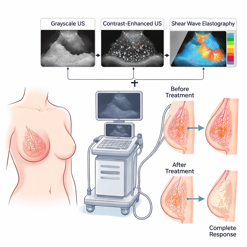
Two advanced ultrasound tools working together
The study focused on contrast-enhanced ultrasound (CEUS) and shear wave elastography (SWE). In CEUS, tiny gas-filled microbubbles are injected into a vein and act as echo‑rich markers of blood flow, outlining the true extent of the tumor and its small vessels in real time. SWE, in contrast, sends gentle sound waves through the breast to measure how much tissues move; very stiff areas, often linked to dense tumor and a rigid supporting scaffold, stand out in color maps. Sixty women with stage II–IV invasive breast cancer had standard ultrasound, CEUS, and SWE scans both before starting chemotherapy and again shortly before surgery. The team recorded the maximum tumor size on regular ultrasound and CEUS, and the maximum stiffness value on SWE, and then calculated how much each of these numbers dropped with treatment.
What the scans revealed about response
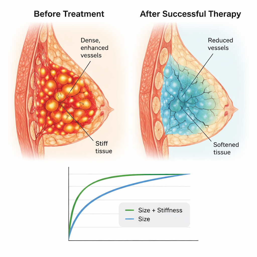
After surgery, pathologists classified each case as either complete response (no cancer cells left in the breast) or non-complete response. Of the 60 women, 28 reached complete response and 32 did not. Before chemotherapy, the two groups looked similar on ultrasound and stiffness measurements. After treatment, however, striking differences emerged. In women who achieved complete response, CEUS often showed no remaining contrast uptake in the tumor area, meaning blood flow had nearly vanished, and SWE maps showed stiffness falling to levels similar to normal breast tissue. On average, tumors in this group shrank by over 90% in size on CEUS and lost about 76% of their stiffness. In the non‑complete response group, size and stiffness also decreased but far less dramatically.
Stronger predictions with a combined signal
The researchers used statistical models to see which measurements best separated complete responders from non-responders. They found that the percentage drop in tumor size on CEUS and the percentage drop in stiffness on SWE were each meaningful on their own. But the best performance came from combining both pieces of information. This paired measure correctly distinguished the two groups much more often than using size change or stiffness change alone. The approach also held up across different biological types of breast cancer, although hormone receptor–negative and certain HER2-related tumors tended to show larger changes, consistent with their known higher sensitivity to chemotherapy.
What this means for patients and care teams
The study suggests that pairing contrast-enhanced ultrasound with shear wave elastography could give doctors a powerful, repeatable way to monitor how a breast tumor is responding to chemotherapy well before surgery takes place. Instead of relying only on whether a lump feels smaller or looks shorter on a basic scan, clinicians could watch for telltale losses in blood supply and stiffness that hint a tumor is truly melting away. While the work was done in a relatively small group and needs confirmation in larger trials, it points toward a future in which a brief, noninvasive imaging session might help tailor treatment in real time—sparing some patients from ineffective regimens and giving others greater confidence that their therapy is on track.
Citation: Wang, Y., Jiang, X., Jiao, Y. et al. The utilization of CEUS and SWE for predicting pathological complete response to neoadjuvant chemotherapy for invasive breast cancer. Sci Rep 16, 7434 (2026). https://doi.org/10.1038/s41598-026-37698-2
Keywords: breast cancer imaging, neoadjuvant chemotherapy, ultrasound, tumor stiffness, treatment response