Clear Sky Science · en
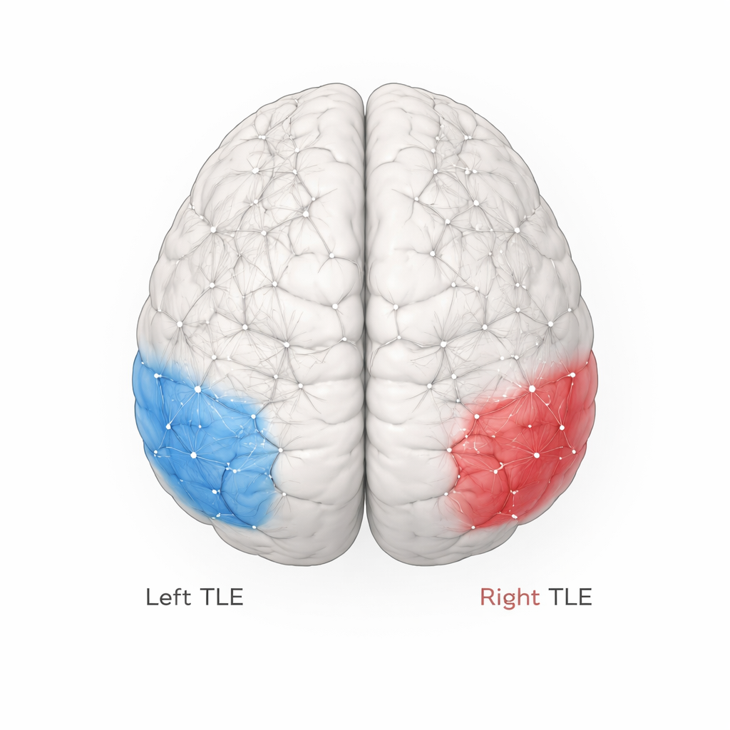
Commonalities and distinctions of static and dynamic functional connectivity density between left and right temporal lobe epilepsy
Why this matters for people living with seizures
Epilepsy is often described as a problem in a single "hot spot" of the brain where seizures begin. This study challenges that simple picture for temporal lobe epilepsy, the most common form of focal epilepsy. Using advanced MRI methods, the researchers show that seizures arising from the left and right temporal lobes disturb wide-ranging brain networks in different ways. These network changes may help explain why people with apparently similar diagnoses can have different memory problems, sensory symptoms, and treatment responses — and they hint at new ways to tailor care.
Looking at the brain’s wiring while it rests
The team scanned 46 people with left temporal lobe epilepsy (LTLE), 43 with right temporal lobe epilepsy (RTLE), and 53 healthy volunteers. Instead of asking participants to do tasks, they recorded brain activity while people simply lay still with eyes closed. They focused on “functional connectivity density,” a measure of how many other areas each tiny spot in the brain talks to on average. They examined this in two ways: as a static snapshot across the whole scan, and as a dynamic measure that captures how these connections rise and fall over time. This allowed them to move beyond traditional approaches that start from a few preselected regions, and instead map communication patterns across the entire brain.
A shared weak point in both forms of temporal lobe epilepsy
Despite differences in where their seizures start, both LTLE and RTLE patients showed a common deficit: reduced connectivity in the temporal lobe on the same side as the epileptic focus. This “lateral temporal lobe” region helps process sounds, language, and complex visual information, and is closely tied to memory and emotion. In everyday terms, it acts like a busy hub that routes information between many other brain areas. Lower connectivity here suggests that this hub has partially lost its central role in the network. Consistent with this, both patient groups performed worse on a standard verbal learning and memory test than healthy volunteers, although the pattern of scores was somewhat worse in the LTLE group.
When seizures start on the left: broader and more symmetric disruption
For people with LTLE, the damage was not confined to the left side. Connectivity density was also reduced in the temporal lobe on the opposite (right) side, and follow-up analyses showed widespread changes in frontal, parietal, and other regions. In other words, left-sided seizures were linked to a more bilateral and extensive reshaping of brain networks. Because the left hemisphere is usually dominant for language in right-handed people, the authors suggest that seizures in this critical language hub may spread more efficiently across pre-existing pathways, disturbing connections in both hemispheres. This broader network disturbance may help account for the more pronounced memory difficulties seen in the LTLE group.
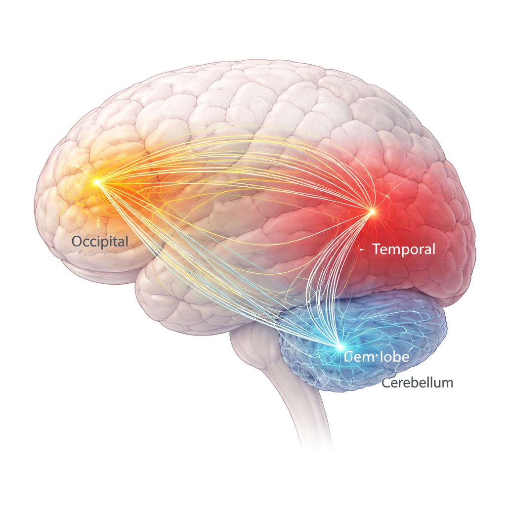
When seizures start on the right: visual and balance centers stand out
Patients with RTLE showed a different signature. Their right temporal lobe connectivity was reduced, as expected, but they also had increased connectivity and greater moment-to-moment variability in parts of the left occipital lobe, a core visual area, along with reduced variability in the left cerebellum, which supports coordination and cognition. Additional analyses confirmed that only the RTLE group showed altered communication between the occipital lobe, cerebellum, and other regions. The authors interpret this pattern as a mix of disturbance and possible compensation: visual areas may become over-connected and unstable as the brain tries to route information around damaged pathways, while the cerebellum may lose some of its usual flexibility in helping the rest of the brain adapt.
What this means for understanding and treating epilepsy
To someone living with temporal lobe epilepsy, these findings reinforce that the condition is not just a small patch of misfiring tissue, but a whole-network disorder whose shape depends on which side of the brain is affected. Left- and right-sided epilepsy share a common weakening of key temporal hubs, yet left-sided disease tends to disturb networks more widely, whereas right-sided disease more often pulls in vision and balance systems. Although the study’s tests of using these network markers to distinguish LTLE from RTLE were promising but not yet strong enough for clinical use, the work shows that combining static and time-varying measures of connectivity can reveal both damage and the brain’s attempts to compensate. In the long run, such network-level insights could guide more personalized predictions of cognitive problems and help refine surgical and non-surgical treatments.
Citation: Song, C., Zhang, X., Cheng, J. et al. Commonalities and distinctions of static and dynamic functional connectivity density between left and right temporal lobe epilepsy. Sci Rep 16, 6652 (2026). https://doi.org/10.1038/s41598-026-37646-0
Keywords: temporal lobe epilepsy, brain networks, functional connectivity, resting-state fMRI, cognitive impairment